Physician-directed clinical protocol

Medical Protocol for Perianal
Hyperpigmentation

An anatomically guided approach to acid selection, controlled application, skin-barrier preservation and clinical follow-up in the perianal region.

01

Surface Affinity

Hydrophilic and lipophilic characteristics of the target area.

02

Epidermal Thickness

Thin transitional tissue versus thicker keratinized skin.

03

Anatomical Safety

Controlled placement and prevention of migration toward the anal canal.

Medical evaluation required Individualized acid selection Anatomically defined treatment boundaries
Clinical anatomy
Clinical anatomical illustration identifying the external anal margin and the surrounding perianal area
Treatment principle

Acid selection and treatment boundaries must be adapted to local epidermal characteristics, surface affinity and proximity to non-keratinized tissue.

Clinical protocol developed by Dr. Alain Tenenbaum & Mauro Tiziani

Clinical Navigation
1.5 Structured Clinical Navigation

PERIANAL TREATMENT PROTOCOL

Clinical Protocol Index

Navigate directly to each clinical phase of the protocol, from anatomical assessment and acid selection to recovery, recurrence prevention and emergency management.

This protocol is intended for qualified healthcare professionals and must always be adapted to individual anatomy, phototype, barrier condition and clinical response.

Clinical decision framework

Clinical Overview

Safe and effective treatment of perianal hyperpigmentation requires more than the selection of a depigmenting acid. The protocol must integrate the physicochemical characteristics of the target surface, local epidermal thickness and the anatomical safety limits of the treatment field.

01 Physicochemical parameter

Surface Affinity

The relative hydrophilic or lipophilic character of the treatment surface influences acid solubility, distribution and penetration.

Hydrophilic Lipophilic
Clinical implication

Acid family and vehicle should be selected according to the characteristics of the target surface rather than according to pigmentation alone.

02 Structural parameter

Epidermal Thickness

Penetration differs between thin transitional epithelium and thicker, more keratinized perianal skin.

Thin Thick
Clinical implication

Concentration, exposure time and number of applications must be adapted to the local resistance of the epidermal barrier.

03 Safety parameter

Anatomical Safety

The treatment field must be defined according to its proximity to the external anal margin, anal canal and non-keratinized epithelium.

Target zone Exclusion zone
Clinical implication

Product migration beyond the planned treatment boundary must be prevented through conservative placement and appropriate protection.

Integrated clinical principle

Acid selection is the result of three combined assessments

No single acid or concentration is suitable for the entire perianal region. Treatment planning must combine surface affinity, epidermal thickness and anatomical safety before defining the formulation, concentration, application boundary and exposure time.

Surface affinity Hydrophilic / Lipophilic
Epidermal thickness Thin / Thick
Anatomical safety Target / Exclusion
Individualized Treatment Strategy
Anatomical mapping before treatment

Anatomical Treatment Zones

The perianal region is not a uniform treatment surface. Safe protocol planning requires a clear distinction between keratinized perianal skin, the external anal margin, the anal canal and the more proximal rectal anatomy.

External anal margin and surrounding perianal skin

The external anal margin forms the immediate visible boundary of the anal opening, while the perianal area extends outward over the surrounding external skin.
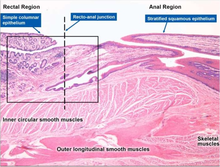
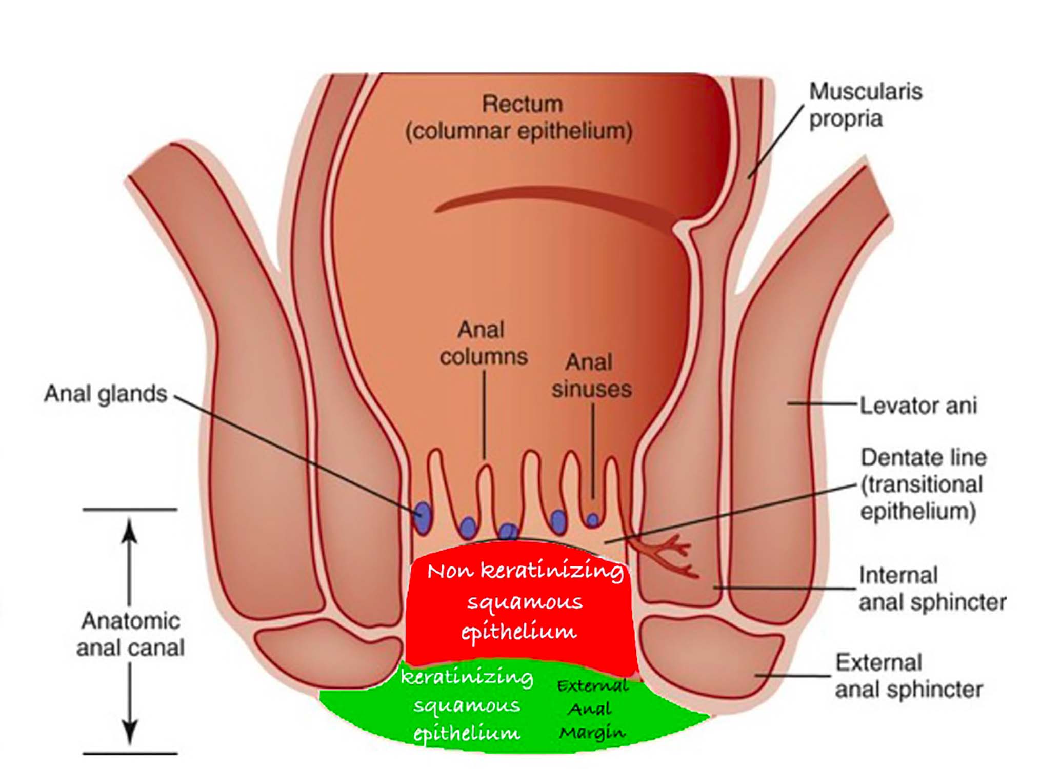
Anatomical illustration distinguishing the external anal margin, anal canal and rectal region View full size
Anatomical overview showing the transition from external perianal skin toward the anal canal and the more proximal rectal region.

External anal margin is not synonymous with perianal skin

The perianal area includes the skin surrounding the anal opening. The external anal margin is the innermost visible external boundary where this skin approaches the opening and transitions toward the specialized epithelium of the anal canal.

Which external zone is closer to the rectum?

The external anal margin is closer to the internal anorectal tract than the surrounding perianal skin. The anal canal, however, lies between the external margin and the rectum.

Treatment-boundary principle

As the application field approaches the anal opening, product placement must become increasingly conservative and protection against uncontrolled migration becomes essential.

Four distinct anatomical levels

Treatment planning must separate external cutaneous targets from internal structures that remain outside the treatment field.
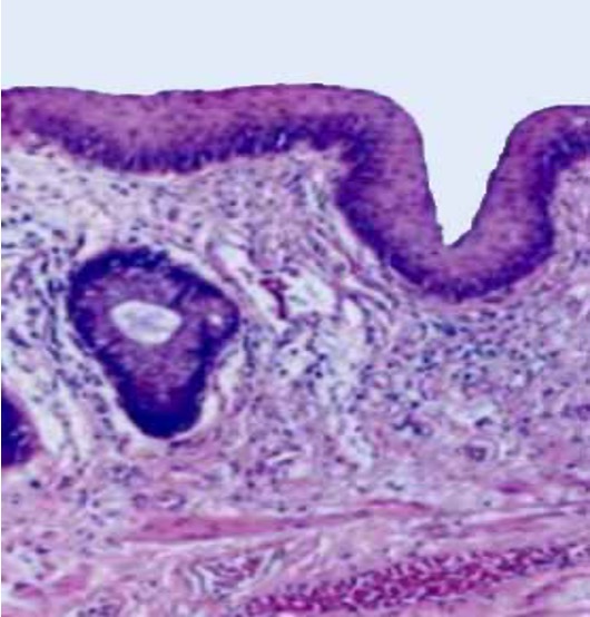
01 External target zone
Histological illustration representing external perianal skin

Perianal Skin

External skin surrounding the anal opening. It presents a more conventional cutaneous surface than the specialized epithelium situated inside the anal canal.
  • Surrounds the anal opening
  • Predominantly external cutaneous surface
  • Variable pigmentation and epidermal thickness
02 High-precision boundary
Clinical safety illustration identifying the external anal margin

External Anal Margin

The immediate external boundary of the anal opening. It requires greater precision because of its direct proximity to the anal canal and transitional epithelium.

  • Innermost visible external boundary
  • Immediately adjacent to the anal canal
  • Increased risk from uncontrolled product migration
03 Exclusion zone

Anal Canal and Anoderm

The anal canal begins internally at the anal verge. Its distal segment includes specialized squamous epithelium that differs from ordinary external skin.
  • Internal anatomical structure
  • Contains highly sensitive specialized epithelium
  • Outside the external cosmetic treatment field
04 Anatomical orientation
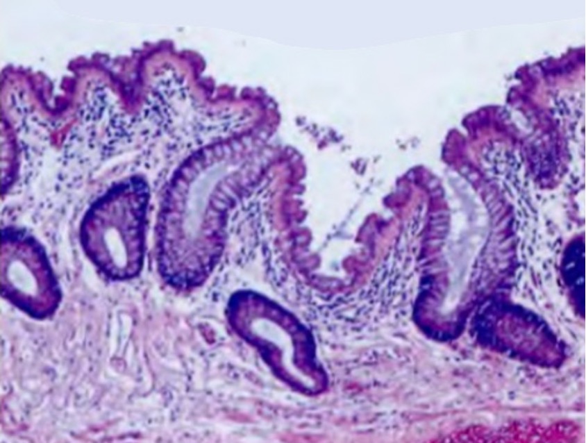
Histological illustration representing rectal anatomy

Rectum

The rectum is a more proximal internal structure located above the anal canal. It is not directly contiguous with external perianal skin.

  • Located proximal to the anal canal
  • Not an external treatment zone
  • Included only for anatomical orientation

Anatomy changes across a very short treatment distance

Surface characteristics and epidermal resistance are not identical throughout the region. These differences must be mapped before an acid-selection decision is made.
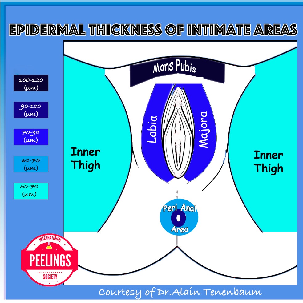
Structural variation

Epidermal Thickness

Diagram comparing thin and thicker epidermal areas within the perianal region View full size

Thin and thicker areas may respond differently to the same formulation, concentration, number of layers and exposure time.
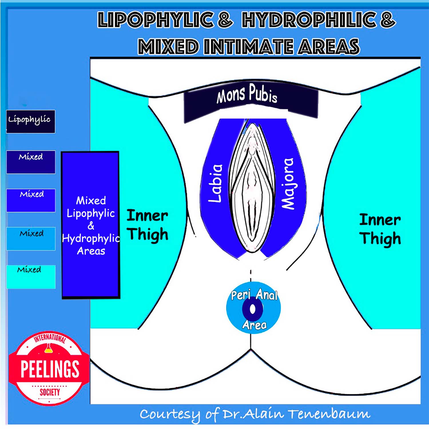
Surface variation

Hydrophilic and Lipophilic Areas

Diagram distinguishing hydrophilic and lipophilic areas within the perianal region View full size

Local moisture, occlusion and lipid characteristics influence product distribution and must be assessed independently from visible pigmentation.

Physician-directed product selection

Acid Selection Framework

Acid selection must be based on the biological and anatomical characteristics of the treatment zone rather than on pigmentation intensity alone. The same formulation must not be applied uniformly across the entire perianal region.
01
Surface behavior Hydrophilic or Lipophilic
02
Barrier resistance Thin or Thick Epidermis
03
Anatomical boundary Target or Exclusion Zone
Individualized Acid Strategy
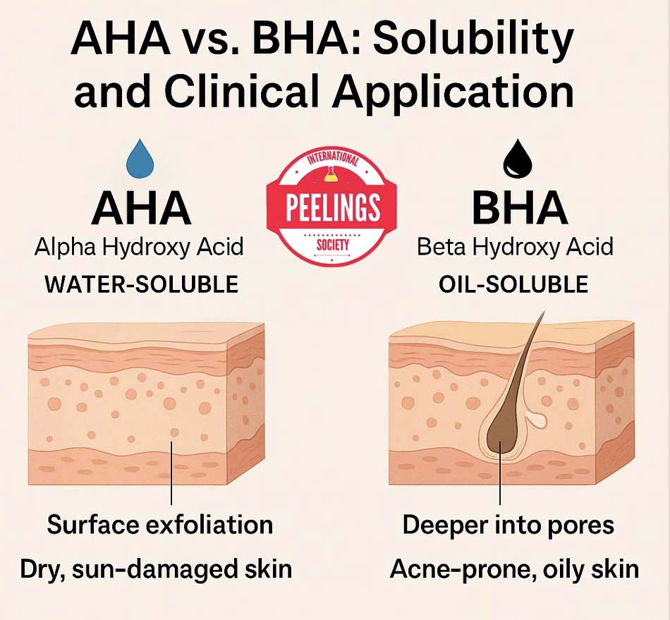
01

Match Acid Solubility to Surface Affinity

Diagram comparing the water solubility of alpha hydroxy acids with the lipid affinity of beta hydroxy acids View full size
Solubility influences product distribution across moist, occluded and lipid-rich cutaneous environments.
Water-associated environment

Hydrophilic Selection

Water-soluble acids are considered when the target surface is relatively hydrated and when uniform superficial distribution is required.

Typical family Alpha Hydroxy Acids
Lipid-associated environment

Lipophilic Selection

Lipid-soluble acids may distribute more effectively across sebaceous, occluded or relatively lipid-rich cutaneous surfaces.

Typical family Beta Hydroxy Acids
Selection principle

Hydrophilic or lipophilic classification does not independently determine treatment suitability. It must be interpreted together with epidermal thickness, anatomical location, formulation, concentration and contact time.

02

Adapt Intensity to Epidermal Resistance

Acid behavior is not defined by concentration alone. Penetration is modified by epidermal thickness, formulation, pH, acid availability, contact time, number of layers, occlusion and local skin condition.

Lower structural resistance

Thin or Transitional Area

Thin tissue requires a conservative approach because a limited application may produce a disproportionately strong biological response.

  • Lower initial intensity
  • Reduced number of layers
  • Shorter observation intervals
  • Strict control of product spread
Intermediate resistance

Standard Cutaneous Area

Conventional external perianal skin may permit gradual treatment, provided that barrier condition and individual response have been assessed.

  • Progressive titration
  • Endpoint-guided application
  • Reassessment between layers
  • Individualized session interval
Greater structural resistance

Thicker Keratinized Area

Thicker keratinized skin may require a different formulation or progressive intensification, but anatomical safety limits remain unchanged.
  • Gradual rather than abrupt escalation
  • Response-based layer adjustment
  • No automatic increase based on color
  • Barrier recovery verified before retreatment
Variables controlling functional penetration
Formulation Free-acid availability Concentration Contact time Number of layers Barrier integrity Local hydration Occlusion
03

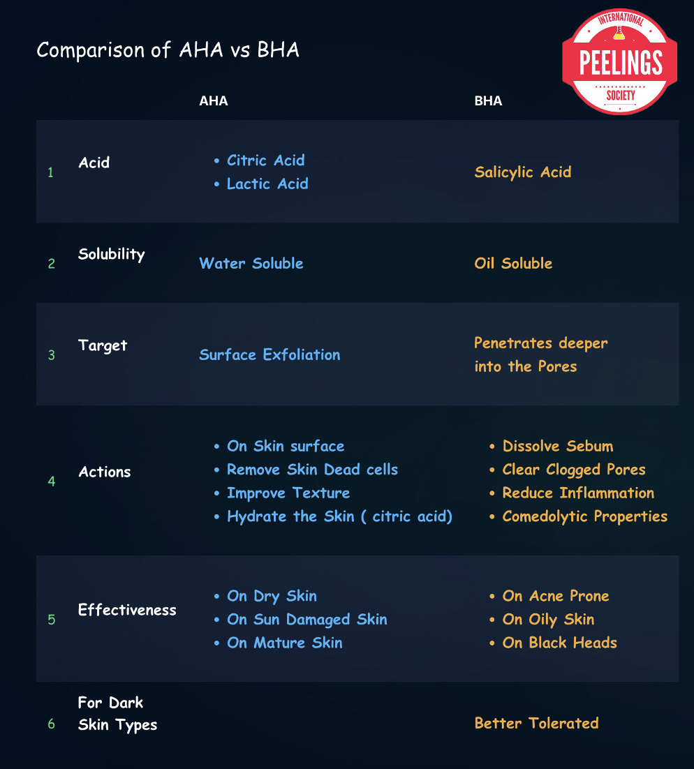
Different Acids Serve Different Clinical Roles

Clinical comparison between alpha hydroxy acids and beta hydroxy acids View full size
AHA
Alpha Hydroxy Acids Predominantly hydrophilic

Considered for controlled superficial action on appropriately selected external cutaneous zones.

Decision basis Hydration, epidermal resistance and tolerance
BHA
Beta Hydroxy Acids Predominantly lipophilic

Considered where lipid affinity is clinically relevant and the selected external zone can be treated safely.

Decision basis Surface affinity, barrier status and formulation
TCA
Trichloroacetic Acid Coagulative acid requiring strict spatial control

TCA is not selected according to hydrophilic or lipophilic affinity. Its use depends on precise anatomical placement, controlled concentration, formulation viscosity and strict exclusion of internal anoderm and anal-canal epithelium.

Critical restriction Use only a validated viscous formulation near the external anal margin
04

Zone-Specific Acid Strategy

The external anal margin and the surrounding perianal skin must not be treated as a single uniform surface. Each zone requires an adapted acid family, formulation viscosity, application method and safety boundary.

High-precision external boundary

External Anal Margin

Controlled placement

This visible external boundary is situated immediately adjacent to the anal opening. Its proximity to the anal canal makes vehicle viscosity and spatial control primary safety parameters.

Surface profile External cutaneous boundary with variable dryness
Main safety issue Run-off or lateral migration toward the anal canal
Primary safety parameter Gel or cream vehicle limiting uncontrolled lateral diffusion
Fundamental safety principle
Vehicle Viscosity Controls Acid Placement

When treatment is performed close to the anal canal, the formulation must remain precisely where it is deposited. A gel or cream vehicle reduces lateral spreading and run-off compared with a freely flowing liquid solution.

In this high-precision boundary zone, the vehicle is not merely a carrier. Its viscosity is an integral component of the safety protocol.

Physician-selected formulation options
  • Alternating acid strategy according to tissue response and the biological objective of each session
  • Salicylic acid in a vehicle selected for controlled placement
  • TCA only in a validated gel or cream formulation when clinically indicated on clearly identified external cutaneous tissue
  • Freely flowing TCA solutions are excluded close to the anal opening because they cannot provide equivalent spatial control
Critical boundary rule

The product must remain on the clearly identified external cutaneous surface. Application into the anal opening, anoderm or anal canal is excluded.

Surrounding external skin

Perianal Area

Broader cutaneous field

The surrounding perianal skin is usually relatively dry because of limited sebaceous activity, although sweat, occlusion, mucus or hygiene-related factors may temporarily increase surface moisture.

Surface profile Dry to variably moist or occluded
Main safety issue Irritation and barrier disruption
Selection priority Match the vehicle to hydration and local tolerance
Physician-selected formulation options
  • AHA or BHA selection according to local surface affinity
  • Salicylic acid may be considered in a compatible vehicle
  • Alcohol-rich vehicles may be irritating in sensitive skin
  • TCA and Jessner-type liquid solutions must not be used indiscriminately across the entire perianal field
Barrier-preservation rule

Product choice must account for sensitivity, hygiene practices, friction, occlusion and the risk of post-inflammatory hyperpigmentation.

External anal margin Precision, viscosity and migration control
Surrounding perianal skin Surface affinity, barrier status and uniformity
05

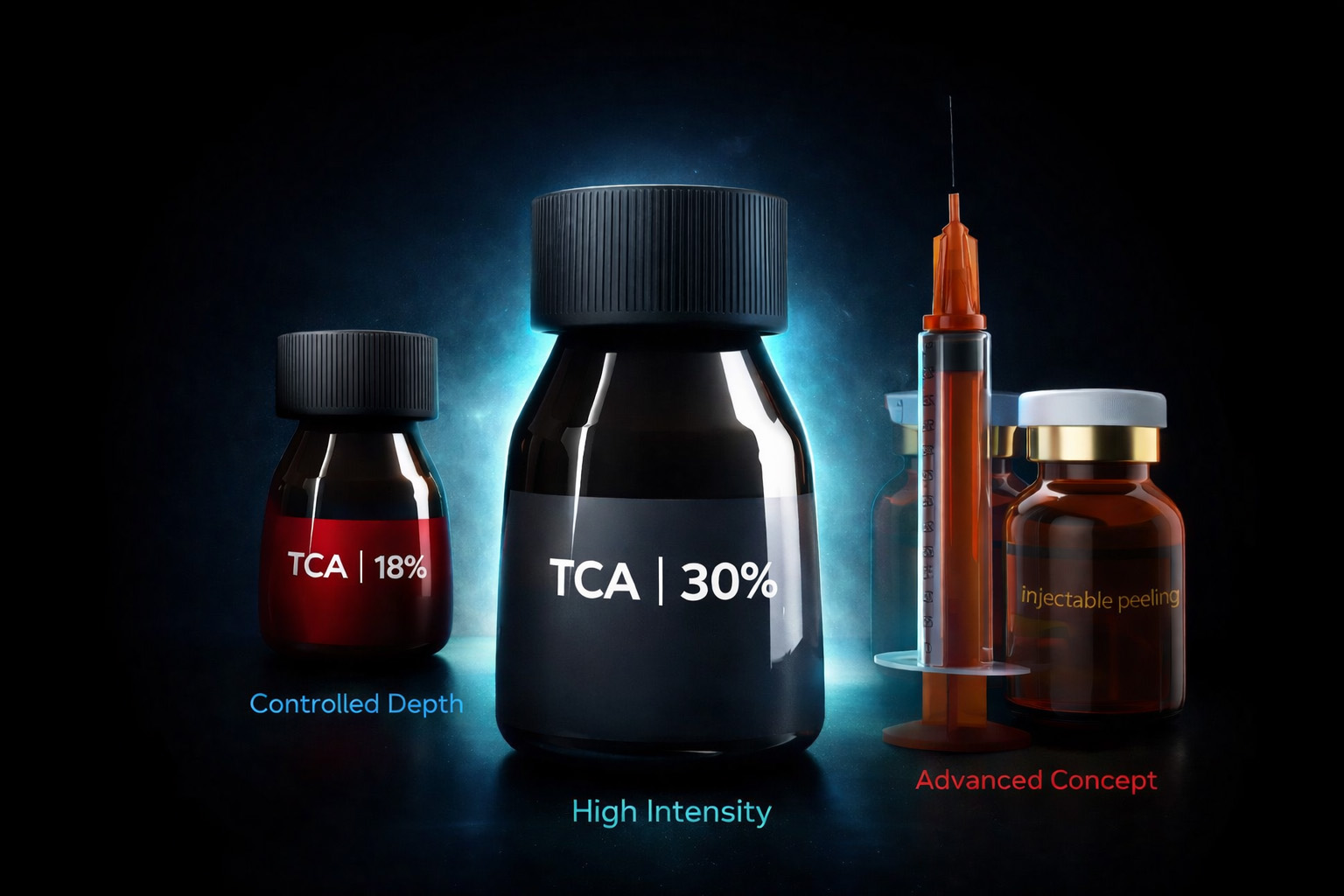
TCA Gel Concentration Has a Validated Stability Limit

Current validated gel platform Maximum Stable TCA Concentration
18% w/w

Within the TCA gel formulation platform used in this protocol, 18% w/w represents the highest validated concentration at which adequate gel stability, viscosity and reproducible spatial application are maintained.

Clinical priority

Spatial Control Takes Priority Over Nominal Concentration

Close to the anal canal, a stable viscous formulation may provide a more appropriate safety profile than a stronger freely flowing solution. Increasing the nominal TCA concentration is not useful if the vehicle can no longer maintain controlled, localized deposition.

Stable viscosity Controlled placement Higher concentration alone
01

Why Use a Gel?

The gel vehicle limits run-off and uncontrolled lateral spreading, permitting more precise deposition on the selected external cutaneous zone.

02

Why Limit the Concentration?

Above the validated 18% w/w concentration, the current formulation does not maintain sufficiently validated gel stability and reproducible rheological behavior.

03

What Must Be Avoided?

A stronger nominal concentration must not be obtained at the expense of vehicle stability, predictable viscosity or anatomical placement control.

Protocol formulation rule

Near the anal canal, select the highest concentration that remains validated as a stable, spatially controllable gel—not the highest TCA percentage that can be prepared as a liquid solution.
06

Concentration Alone Does Not Define Treatment Intensity

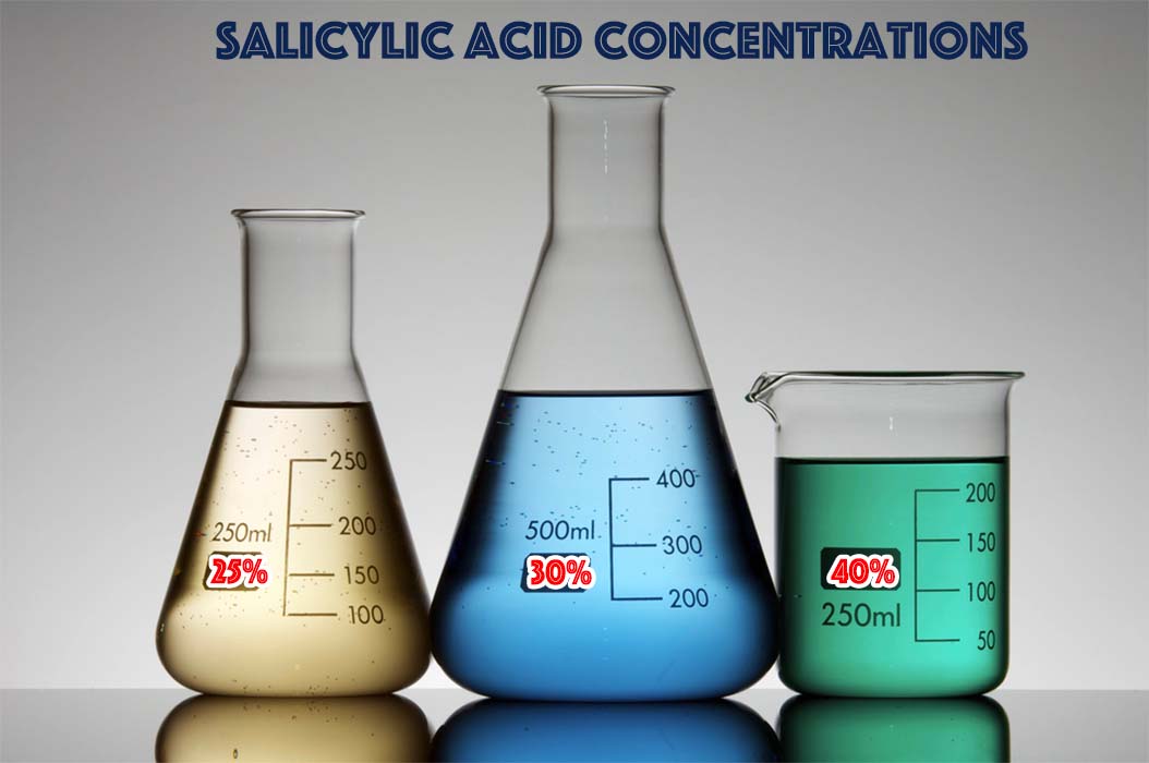
Comparison of three salicylic acid concentrations View full size

A percentage is only one component of the formulation

Products displaying the same named acid may behave differently according to their vehicle, pH, free-acid fraction, viscosity, application technique and treated anatomy.

Concentration Predictable depth
  • Do not compare products by percentage alone.
  • Do not extrapolate facial protocols directly to the perianal region.
  • Do not increase intensity solely because pigmentation appears darker.
  • Observe the tissue response after each controlled application.

Select the Zone Before Selecting the Acid

Anatomical classification precedes product choice. The acid is selected only after the treatment field and its exclusion boundaries have been defined.

Step 01 Identify the Anatomical Zone

Separate external perianal skin from the external anal margin and internal exclusion zones.

Step 02 Assess Surface Affinity

Determine whether the local surface behaves predominantly as a hydrophilic or lipophilic environment.

Step 03 Estimate Barrier Resistance

Evaluate epidermal thickness, hydration, irritation and previous treatment response.

Step 04 Select and Titrate

Define the acid family, formulation, initial intensity, number of layers and observation interval.

Clinical preparation before acid application

Pretreatment Protocol

Pretreatment determines whether the selected acid can be applied safely, precisely and reproducibly. Medical screening, anatomical mapping, baseline documentation and preparation of a clean, dry and clearly delimited treatment field must be completed before the first layer is applied.

Core safety principle

Prepare the Patient, the Anatomy and the Treatment Field

Acid selection alone does not create a safe protocol. The treatment field must be medically appropriate, anatomically mapped, microbiologically controlled, completely dry and protected against unintended product migration.

01

Medical Assessment and Baseline Documentation

Medical consultation

Confirm Treatment Eligibility

Review the medical history, previous peel response, current medications, allergies, healing capacity and any history of post-inflammatory hyperpigmentation.

Local examination

Exclude Active Disease

Inspect for irritation, dermatitis, infection, fissures, ulceration, bleeding, inflammatory lesions or anorectal disease requiring evaluation before cosmetic treatment.

Pigmentary risk

Record the Skin Phototype

Document the Fitzpatrick phototype, baseline pigmentation, surrounding color variation and the risk of inflammatory hyperpigmentation after treatment.

Patient authorization

Obtain Informed Consent

Explain expected benefits, limitations, discomfort, recovery, pigmentation variability, aftercare requirements and the possibility that several sessions may be required.

Baseline photography

Standardize the Clinical Documentation

Same camera Same lighting Same distance Same patient position No image filters Date recorded

Photographs should document the baseline pigmentation and treatment boundaries before cleansing or product application. Consistent photographic conditions are essential for interpreting subsequent clinical change.

02

Define the Treatment and Exclusion Zones

01

Identify the Perianal Cutaneous Field

Delimit the surrounding external skin that may be included in the depigmentation protocol.

02

Identify the External Anal Margin

Recognize the high-precision boundary immediately adjacent to the anal opening.

03

Mark the Internal Exclusion Zone

The anal canal, anoderm and internal mucosal structures remain outside the external depigmentation treatment field.

04

Adapt the Safety Boundary

Increase the untreated safety margin when using a formulation with lower viscosity or greater potential for lateral spread.
03

Cleanse, Control the Microbial Environment and Dry the Field

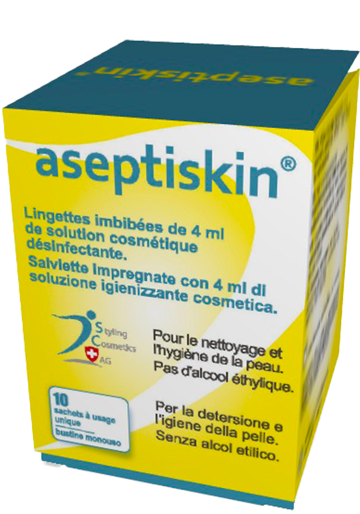
Aseptiskin cleansing and antiseptic preparation product
Pretreatment product Aseptiskin View product information
Mandatory field preparation

Aseptiskin Before Every Peel Application

The treatment area must be carefully cleansed before acid application. Aseptiskin is used to reduce surface contamination, remove residues and prepare a controlled treatment field without replacing the need for precise anatomical protection.

Step 01 Remove Surface Residues

Eliminate cosmetics, creams, perspiration, mucus, lubricant residues and visible contamination.

Step 02 Apply Aseptiskin

Prepare the complete external treatment field using a clean and controlled technique.

Step 03 Allow Complete Drying

Do not apply an acid over a wet or incompletely dried preparation layer.

Learn More About Aseptiskin
04

Protect the Anal Opening and Adjacent Sensitive Tissue

A

Confirm Complete Dryness

Protection materials and surrounding tissue must be dry so that the barrier remains stable throughout the application.

B

Protect the Exclusion Boundary

Apply an appropriate protective barrier to tissue that must remain outside the treatment field.

C

Control Patient Position

Maintain a stable position that permits visualization of the field and reduces the risk of acid run-off or unintended contact.

D

Prepare Immediate Removal Materials

Clean gauze, appropriate removal materials and the planned neutralization method, when relevant, must be immediately accessible.

Vehicle-specific protection

The Lower the Viscosity, the Wider the Required Safety Margin

Gel and cream vehicles provide greater spatial control than freely flowing solutions. Nevertheless, viscosity does not replace anatomical protection, direct visualization or conservative acid loading.

05

Confirm the Acid, Vehicle and Initial Application Strategy

Review Before Treatment

These resources provide additional information on pretreatment cleansing and the clinical differences between salicylic acid and TCA.

Physician-controlled, zone-specific sequence

Five-Step Biological
Treatment Architecture

Perianal hyperpigmentation must not be treated as a uniform surface. The protocol combines controlled direct-acid stimulation, frosting modulation, targeted depigmentation, hydration and final penetration enhancement within three anatomically distinct treatment fields.

05 Functional steps
03 Treatment fields
01 Physician-controlled architecture
A
Spatial treatment model

Three Concentric Treatment Fields

The original lesion, peri-wound and surrounding-skin model is adapted here to perianal hyperpigmentation. Each field receives a distinct acid intensity and post-frosting sequence.

01 Highest pigment target

Central Hyperpigmented Treatment Zone

The most intensely pigmented external cutaneous field. When a frosting-based step is selected, the endpoint may be allowed to become completely white and to cover the mapped target.

  • Use the highest acid intensity selected for the session.
  • Allow complete white frosting when clinically intended.
  • Do not apply Peeling de Luxe Plus during active white frosting.
  • Wait for defrosting before applying Peeling de Luxe Plus.
02 Precision transition zone

Narrow Peripheral Transition Zone

The narrow external ring immediately surrounding the central target. A lower acid concentration is used, although white frosting may still appear and must then be stopped without delay.

  • Use a lower concentration than in the central target.
  • Observe continuously for the first sign of white frosting.
  • Apply Peeling de Luxe Plus immediately if frosting becomes white.
  • Apply StretchPeel directly after the frosting stopper.
03 Hydration and transition field

Larger Surrounding Cutaneous Area

The broader surrounding skin receives a low-intensity approach designed to avoid frosting and maintain a continuous hydration gradient between treated and untreated skin.

  • Use only a low-concentration acid when indicated.
  • Avoid frosting in this larger surrounding field.
  • Peeling de Luxe Plus is generally unnecessary here.
  • Apply Les Félins to preserve non-greasy hydration.
B
Sequential biological control

The Complete Five-Step Sequence

Each step has one dominant biological function. The architecture progresses from controlled stimulation to endpoint regulation, pigment modulation, hydration and final enhancement of penetration.

01
Direct acid

Direct Acid Application

Primary biological trigger

Salicylic acid in ethanol, another selected salicylic formulation or a TCA gel initiates the controlled biological response. Acid, concentration and vehicle are selected independently for each field.

Central zone Controlled complete endpoint

White frosting may be deliberately obtained over the entire mapped hyperpigmented target when clinically appropriate.

Transition zone Reduced concentration

Use less acid than centrally and prepare to stop white frosting immediately if it develops.

Surrounding zone Low-intensity field

The objective is a conservative biological stimulus without frosting or unnecessary extension of injury.
Vehicle control: Close to the anal canal, prefer a formulation with sufficient viscosity to prevent migration beyond the mapped external field.
02
Frosting stopper

Peeling de Luxe Plus

Frosting modulation technology
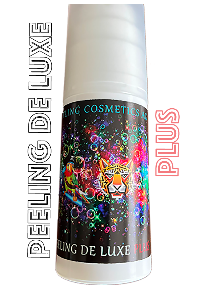
Peeling de Luxe Plus frosting stopper
Retinoic-acid–structured AHA system
Product information

Peeling de Luxe Plus is not presented here as a conventional metabolic peel alone. Within this protocol it functions as a frosting stopper and epidermal modulator, increasing physician control and preserving a wider safety margin.

Central target
Wait for defrosting

Do not cover active white frosting. Allow the white endpoint to defrost, then apply Peeling de Luxe Plus over the central lesion.

Narrow transition zone
Stop white frosting immediately

When the lower-concentration peripheral application becomes white, apply Peeling de Luxe Plus without delay.

Surrounding field
Usually unnecessary

No frosting is sought in the larger surrounding area; therefore, the frosting stopper is generally not required.
Specific salicylic sequence

Peeling de Luxe Plus is applied after white pseudo-frosting produced by salicylic acid in ethanol.

03
Depigmenting cream

StretchPeel

Targeted pigment modulation
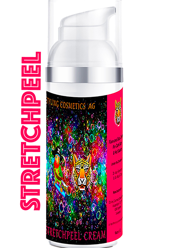
StretchPeel depigmenting cream containing kojic acid
Kojic-acid depigmenting formulation
Product information

StretchPeel is a depigmenting cream containing kojic acid. It continues pigment modulation after the direct-acid phase and after frosting has been brought under control.
  1. Central zone Apply over Peeling de Luxe Plus after complete defrosting.
  2. Narrow transition zone Apply immediately after Peeling de Luxe Plus has stopped white frosting.
  3. Gray frosting phase Reapply StretchPeel once the frosting changes from white to gray.
04
Moisturizing cream

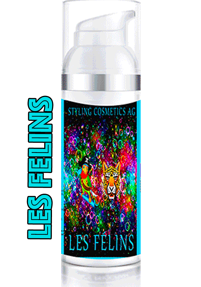
Les Félins

Barrier recovery and moisture equilibrium
Les Félins moisturizing cream
Fast-absorbing, non-greasy moisturizer
Product information

Les Félins is applied only over the larger surrounding cutaneous area. It supports epidermal comfort and creates a continuous hydration field around the more intensively treated central zones.

01Hydration

Maintains moisture in treated and untreated surrounding skin.

02Comfort

Supports tissue comfort after the controlled chemical stimulus.

03Finish

Non-greasy and rapidly absorbed for practical post-procedure use.
05
Penetration enhancer

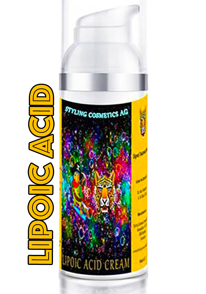
Lipoic Acid

Dual penetration enhancement technology
Lipoic Acid penetration enhancer
Final protocol-enhancing step
Product information

Lipoic Acid is applied after the Peeling de Luxe Plus, StretchPeel and Les Félins sequence. It is not merely another acid: its primary role is to enhance penetration of the formulations already placed on the skin.

A
Direct-acid depth
Deeper Biological Diffusion Without Automatically Increasing Concentration

By enhancing penetration, Lipoic Acid may allow a lower-concentration direct acid to reach a greater biological depth. This can reduce the need to raise acid concentration solely to obtain deeper action.

Lower-concentration acid Enhanced diffusion Greater biological depth
B
Post-peel formulations
Enhanced Penetration of Depigmenting and Moisturizing Products

As the final application, Lipoic Acid also promotes penetration of the previously applied StretchPeel depigmenting cream and Les Félins moisturizing cream.

StretchPeel + Les Félins Lipoic Acid Enhanced penetration
Final functional interpretation

Lipoic Acid therefore serves two complementary purposes: it supports deeper acid action without necessarily increasing acid concentration, and it enhances penetration of the depigmenting and moisturizing formulations already applied.

C
Visual endpoint interpretation

Frosting Control and Post-Frosting Sequence

Frosting is interpreted according to location, intensity, vehicle and evolution over time. The same visual endpoint has a different meaning in the central target and in the narrow peripheral zone.

White pseudo-frosting after salicylic acid in ethanol
White pseudo-frosting after salicylic acid in ethanol during a subsequent treatment session.
White endpoint

Active Frosting Phase

In the central target, wait for defrosting. In the narrow transition zone, stop white frosting immediately with Peeling de Luxe Plus.

Gray endpoint

Post-Frosting Transition

When the frosting becomes gray, reapply StretchPeel to reinforce the depigmenting sequence.

No frosting

Surrounding Hydration Field

The larger surrounding field is managed conservatively and receives Les Félins rather than a frosting-control sequence.

D
Protocol-integrated products

Therapeutic Arsenal

The protocol combines metabolic formulations with physician-selected direct acids. Each product is assigned a specific functional role.
01
Functional formulations

Metabolic and Support Products

Peeling de Luxe PlusFrosting stopper · epidermal modulation
StretchPeelKojic-acid depigmenting cream
Les FélinsMoisturizing and barrier-support cream
Lipoic AcidDual penetration enhancer
02
Primary chemical stimulus

Direct Acids

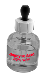
Salicylic acid formulation
BHA family
Salicylic Acid Formulations

Includes salicylic acid in ethanol when controlled white pseudo-frosting is clinically selected.
TCA formulation
Controlled coagulative acid
TCA Formulations

Selected according to anatomical field, concentration, viscosity, endpoint and physician experience.

Explore Direct Acids
Clinical governance

Document Every Zone, Product and Endpoint

Record the selected acid, vehicle, concentration, number of layers, observed endpoint, frosting evolution, timing of each post-frosting product and the precise anatomical limits treated during the session.

01Pre-treatment mapping

Photograph and delineate the three cutaneous fields.

02Endpoint timing

Record whitening, defrosting and gray-frosting transitions.

03Product sequence

Document every application and reapplication in order.

04Follow-up imaging

Use standardized photographs to assess progressive response.

R Recovery architecture

RECOVERY • FOLLOW-UP • SAFETY

Recovery, Long-Term Follow-Up & Clinical Safety

Recovery is not a secondary phase of treatment. Barrier preservation, structured follow-up, controlled home care and early recognition of adverse signs are integral components of the therapeutic protocol.

R.1

EARLY CLINICAL EVOLUTION

Recovery Timeline

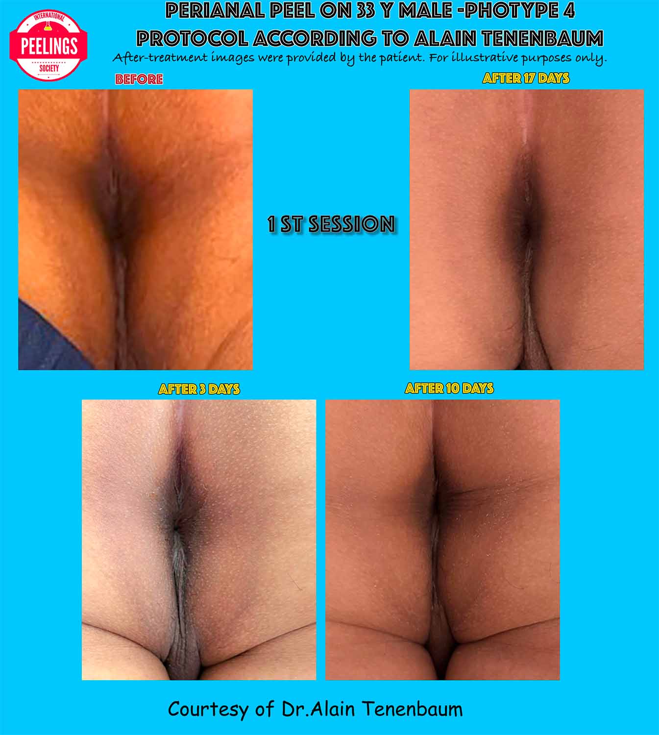
Clinical evolution from day 0 to day 17 after the first perianal peeling session
First-session clinical evolution from the immediate endpoint to biological stabilization.
D0
IMMEDIATE ENDPOINT

Controlled Treatment Response

The physician documents the treated zones, frosting or pseudo-frosting pattern and the immediate cutaneous response.

D3
EARLY RECOVERY

Controlled Desquamation

Transient dryness, mild desquamation or color transition may appear. Friction, perfumes and non-essential cosmetics remain excluded.

D10
BARRIER REORGANIZATION

Progressive Recovery

Epidermal comfort and surface continuity should improve while the depigmenting sequence and hydration strategy are maintained.

D17
BIOLOGICAL STABILIZATION

Clinical Reassessment

Pigment response, tissue tolerance and readiness for the next session are assessed before any further treatment decision.

R.2

HOME RECOVERY PROTOCOL

Recovery Products & Scientific Support

MOISTURE EQUILIBRIUM
Les Félins moisturizing cream

Les Félins

Fast-absorbing, non-greasy moisturizing cream used to maintain comfort and hydration within the larger surrounding cutaneous area.

Scientific information
PIGMENT MODULATION
StretchPeel depigmenting cream

StretchPeel

Kojic-acid depigmenting cream designed to maintain targeted pigment modulation during the post-peel recovery sequence.

Scientific information
PENETRATION ENHANCEMENT
Lipoic Acid penetration enhancer

Lipoic Acid

Final penetration-enhancing step supporting deeper diffusion of the previously applied depigmenting and moisturizing formulations.

Scientific information
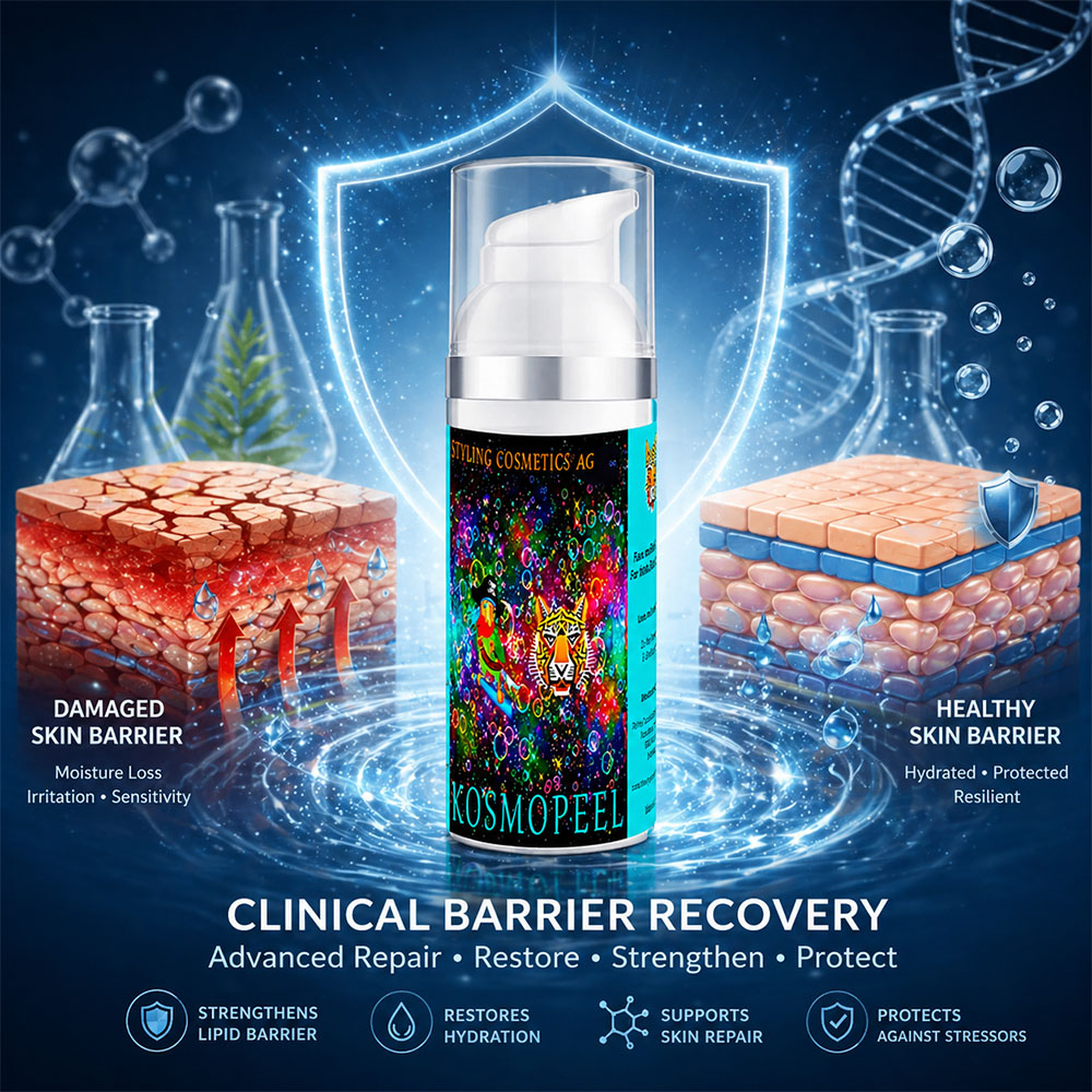
BARRIER SCIENCE
Post-peel barrier instability and corneocyte disruption

Barrier Recovery Science

Clinical guidance on restoring epidermal barrier integrity, maintaining hydration and supporting biological recovery following controlled chemical peeling.

Open scientific guide
R.3

IMMEDIATE POST-PEEL RESTRICTIONS

Protect the Recovering Epidermis

During the early recovery phase, unnecessary chemical exposure, friction and cosmetic interference should be avoided until the epidermal barrier has recovered sufficiently.

No direct sun exposure
01

No Direct Sun Exposure

UV exposure may stimulate melanogenesis and increase the risk of recurrent or post-inflammatory hyperpigmentation.

Biological photoprotection

No perfumes
02

No Perfumes

Fragrances and alcohol-containing perfumed products may irritate the recovering epidermis and interfere with barrier restoration.
No non-essential cosmetics
03

No Non-Essential Cosmetics

Only products specifically recommended within the treatment protocol should be applied to the treated area during recovery.

04

Toilet Hygiene

Avoid direct contact with toilet seats recently disinfected with alcohol. Residual alcohol may irritate treated perianal skin and delay optimal barrier recovery.

R.4

CLINICAL SAFETY CENTER

Rapid Access to Safety Resources

Emergency Skin Rash Kit
EMERGENCY RESOURCE

Emergency Skin Rash Kit

Immediate access to practical guidance for unexpected inflammatory reactions, skin rash or acute post-procedure intolerance.

Open emergency kit
!
RISK ASSESSMENT

Safety & Clinical Risk

Review clinical warning signs, practitioner responsibilities and escalation pathways when recovery does not follow the expected course.

Open safety documentation
×
PRE-TREATMENT CONTROL

Contraindications

Reassess contraindications before every subsequent session, particularly after any unexpected reaction or delayed recovery.

Review contraindications
R.5

LONG-TERM CLINICAL EVOLUTION

Seven-Month Follow-Up

7-MONTH FOLLOW-UP
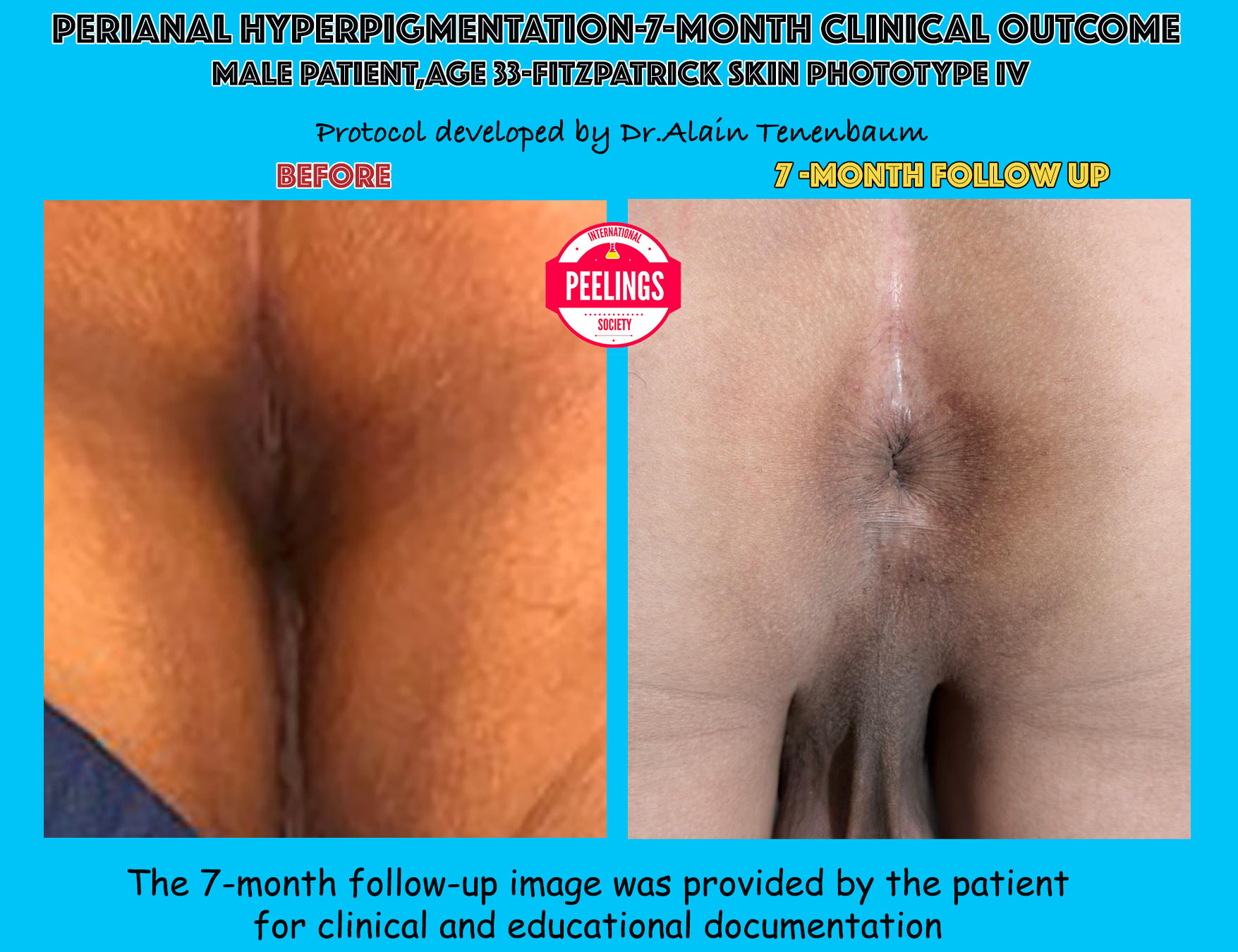
Perianal hyperpigmentation before treatment and after seven months
Long-term clinical evolution after a structured multi-session protocol. Individual results vary according to phototype, pigment depth, biological healing response and adherence to follow-up instructions.
R.6

SCIENTIFIC RESOURCES & CLINICAL SUPPORT

Continue the Clinical Pathway

FINAL CLINICAL STATEMENT

Successful treatment of perianal hyperpigmentation depends not only on appropriate acid selection, but equally on controlled recovery, barrier preservation, structured follow-up and long-term patient compliance.

These complementary biological phases are integral components of the therapeutic protocol rather than separate post-treatment recommendations.

P Patient reassurance protocol

TEMPORARY POST-TREATMENT DARKENING

Don’t Panic

A transient increase in visible pigmentation may occur after the first three treatment sessions. This temporary phase can appear more intense than the original hyperchromia, but it does not automatically indicate treatment failure.

PATIENT MESSAGE

Temporary darkening can be part of the expected biological recovery sequence.

BIOLOGICAL EXPLANATION

Why the Treated Area May Look Darker Before It Improves

During the early phase of the first three treatment sessions, the treated pigmentation may become visually darker, more contrasted or apparently more extensive than before treatment.

01
TRANSIENT RESPONSE

Temporary Pigment Concentration

Pigment may appear more concentrated while the superficial epidermal layers reorganize and the treated chromatic field evolves.

02
RECOVERY PHASE

Inflammatory Color Transition

Mild post-treatment inflammation, dryness and desquamation can temporarily increase visual contrast before barrier recovery is complete.

03
CLINICAL INTERPRETATION

Not Automatically a Treatment Failure

The result should be interpreted within the complete recovery timeline and should not be judged only during the first days following treatment.

Temporary darkening should nevertheless be distinguished from an adverse reaction. Contact the physician when it is associated with significant pain, progressive ulceration, marked swelling, persistent discharge, extensive blistering or another unexpected clinical sign.

EXPECTED VISUAL EVOLUTION

From Temporary Darkening to Clinical Improvement

This timeline is a simplified patient-facing representation. The duration of each phase varies according to phototype, treatment depth, pigment biology and individual healing response.

01 TREATMENT

Controlled Chemical Stimulus

02 FIRST THREE SESSIONS

Temporary Darkening

03 RECOVERY

Barrier Reorganization

04 REMODELING

Pigment Evolution

05 FOLLOW-UP

Clinical Improvement

PATIENT INFORMATION

Explain the Temporary Worsening Before It Occurs

Providing clear information before treatment reduces unnecessary anxiety when the treated hyperchromia appears temporarily darker during the recovery phase.

Patient Information Leaflet
FOR THE PATIENT

Observe, Protect, Document

  • Do not scratch, rub or manually remove desquamating skin.
  • Apply only the products prescribed within the protocol.
  • Avoid perfumes, non-essential cosmetics and direct UV exposure.
  • Send follow-up photographs according to the agreed schedule.
FOR THE PHYSICIAN

Reassure, Compare, Reassess

  • Compare photographs with the immediate post-treatment baseline.
  • Differentiate expected temporary darkening from an adverse inflammatory reaction.
  • Assess barrier recovery before considering another session.
  • Escalate clinical review whenever warning signs are present.

KEY MESSAGE

A darker appearance during early recovery can be temporary. The treatment outcome should be evaluated through serial follow-up, not from a single early post-treatment image.
Documented clinical follow-up

Long-Term Clinical Outcome

Baseline appearance compared with the clinical result documented approximately seven months after initiation of the perianal depigmentation protocol.

7-Month Follow-Up
Clinical comparison showing the perianal area before treatment and approximately seven months after the depigmentation protocol
Baseline, left; seven-month follow-up, right. The long-term follow-up photograph was voluntarily provided by the patient for clinical and educational documentation.
01

Visible Pigmentation Reduction

The follow-up appearance demonstrates a sustained reduction in visible perianal hyperpigmentation.

02

Improved Color Homogeneity

A more homogeneous transition can be observed between the treated area and the surrounding skin.

03

Long-Term Result

The clinical improvement remained visible approximately seven months after treatment initiation.

Clinical Interpretation

This individual case illustrates a persistent improvement in visible pigmentation over extended follow-up. The result should be interpreted as a documented individual clinical outcome and not as a guarantee of response in every patient.

M Long-term clinical strategy

MAINTENANCE • MONITORING • RECURRENCE CONTROL

Long-Term Maintenance & Recurrence Control

Maintenance begins only after the active treatment phase has ended, epidermal recovery is complete and the pigment response has become clinically stable.

M.1

TRANSITION FROM ACTIVE TREATMENT

When Does Maintenance Begin?

Maintenance is not an automatic continuation of repeated peeling. It begins only when the physician confirms that the active treatment objectives have been reached and that the treated tissues have entered a stable phase.

The transition to maintenance is a clinical decision based on biological recovery, photographic comparison and pigment stability.

01 PIGMENT RESPONSE

Stable Clinical Improvement

The treated hyperchromia shows a documented and sustained reduction without progressive rebound.

02 BARRIER STATUS

Complete Epidermal Recovery

Desquamation, irritation, dryness and inflammatory signs have resolved.

03 CLINICAL TOLERANCE

No Active Inflammation

The area is comfortable, stable and free from persistent erythema, pain or abnormal sensitivity.

04 DOCUMENTATION

Photographic Confirmation

Standardized images confirm that the improvement is maintained between follow-up visits.

M.2

STRUCTURED LONG-TERM MONITORING

Scheduled Clinical Reassessment

Follow-up intervals should be adapted to the initial pigment depth, phototype, recurrence tendency, tissue tolerance and patient adherence. The timeline below provides a structured framework rather than a fixed universal schedule.

1 MONTH

Early Stability Review

Confirm barrier recovery, compare standardized photographs and identify any early pigment rebound.

3 MONTHS

Intermediate Pigment Assessment

Evaluate whether the clinical improvement remains stable without further active treatment.

6 MONTHS

Recurrence Screening

Identify localized recurrence, progressive darkening or changes in the surrounding cutaneous field.

12 MONTHS

Long-Term Outcome Review

Reassess pigment stability, maintenance needs and the indication for any future treatment cycle.

M.3

CLINICAL DECISION TRIGGERS

When May Maintenance Treatment Be Considered?

DOCUMENTED RECURRENCE

Retreatment Is Based on Evolution, Not on a Fixed Calendar

A new treatment session should not be scheduled automatically. It may be considered when serial clinical evaluation demonstrates a meaningful recurrence and the epidermal barrier is fully stable.

01

Progressive Recurrence

Gradual reappearance of pigmentation confirmed across serial photographs.

02

Localized Pigment Return

Re-emergence of a defined hyperpigmented area after a period of stability.

03

Increasing Contrast

Documented increase in contrast between the central zone and the surrounding skin.

04

Stable Cutaneous Barrier

No active irritation, desquamation, pain or inflammatory reaction is present.

M.4

TREATMENT DEFERRAL

When Not to Retreat

×

Persistent Irritation

Retreatment should be deferred while erythema, burning, sensitivity or discomfort remains present.

×

Unresolved Desquamation

Active scaling, crusting or incomplete barrier recovery indicates that the skin is not ready for another chemical stimulus.

×

Unstable Dyschromia

Rapidly changing pigmentation should be reassessed before any maintenance procedure is considered.

×

Inadequate Documentation

A new session should not be based solely on memory or subjective perception without comparable clinical photographs.

×

Poor Protocol Adherence

Repeated non-adherence to recovery instructions increases risk and reduces the reliability of treatment assessment.

×

Unexpected Clinical Signs

Pain, ulceration, discharge, swelling or abnormal healing require clinical evaluation rather than routine maintenance treatment.

M.5

RECURRENCE PREVENTION

Preserve the Result Without Over-Treating

01
BARRIER PRESERVATION

Maintain Cutaneous Stability

Continue an appropriate moisturizing and barrier-support strategy when clinically indicated.

02
IRRITANT REDUCTION

Avoid Repeated Chemical Irritation

Perfumed products, aggressive cleansing and unnecessary topical agents may contribute to recurrent inflammation.

03
PHOTOPROTECTION

Maintain Biological Photoprotection

Photoprotection remains relevant to long-term pigment control, even when the anatomical area is not routinely sun-exposed.

04
CLINICAL MONITORING

Detect Recurrence Early

Standardized photographs allow subtle pigment changes to be recognized before a major recurrence develops.

MAINTENANCE PRINCIPLE

Maintenance should preserve a stable result, not expose recovered skin to unnecessary repeated treatment.

The indication for retreatment must remain individualized, documented and dependent on recurrence, barrier integrity and clinical tolerance.

P Recurrence prevention strategy

PREVENTION • BARRIER PRESERVATION • PIGMENT CONTROL

Prevention of Recurrent Hyperpigmentation

Long-term pigment control depends on reducing repeated irritation, preserving epidermal barrier stability and identifying individual biological or behavioral triggers that may reactivate hyperchromia.

P.1

CORE PREVENTION PRINCIPLES

Protect the Result Without Over-Treating

01 GENTLE CLEANSING

Use Skin-Friendly Products

Use mild, non-stripping cleansing products and avoid aggressive washing, abrasive exfoliation or repeated mechanical friction.

02 BARRIER PRESERVATION

Maintain Hydration and Comfort

Maintain appropriate hydration and barrier support when clinically indicated, especially after irritation, friction or recurrent dryness.

03 IRRITANT REDUCTION

Avoid Alcohol, Perfume and Abrasives

Fragrances, alcohol-containing products, harsh cosmetics and repeated exfoliation may reactivate inflammation and contribute to pigment recurrence.

04 BEHAVIORAL CONTROL

Do Not Scratch or Pick the Skin

Scratching, picking, rubbing and manual removal of desquamating skin may provoke post-inflammatory hyperpigmentation.

P.2

INDIVIDUAL RECURRENCE TRIGGERS

Identify What May Reactivate Pigmentation

PERSONALIZED PREVENTION

Recurrence Is Not Driven by a Single Universal Cause

Hormonal changes, medication exposure, repeated irritation, friction, inflammation and barrier instability may contribute differently from one patient to another. Prevention should therefore remain individualized.

01

Hormonal Changes

Pregnancy, contraceptive changes or hormone therapy may influence pigment behavior and should be discussed when clinically relevant.

02

Medication-Related Changes

New or modified medication should be reviewed when pigment behavior changes unexpectedly.

03

Repeated Friction

Tight clothing, persistent rubbing or repeated mechanical irritation may sustain local inflammatory signaling.

04

Barrier Instability

Recurrent dryness, irritation or incomplete recovery may increase the risk of renewed hyperchromia.

P.3

WHAT TO AVOID

Prevent Irritation and Unsupervised Treatment

×

Unsupervised Strong Depigmenting Agents

Avoid unsupervised use of hydroquinone, strong corticosteroids or other potent medical agents without appropriate clinical assessment.

×

Aggressive Exfoliation

Avoid scrubs, abrasive devices, repeated acid application or over-exfoliation that may destabilize the epidermal barrier.

×

Perfumed or Alcohol-Based Products

Avoid products likely to irritate the treated anatomical area or trigger recurrent inflammation.

×

Automatic Maintenance Peeling

Do not repeat treatment on a fixed calendar without documented recurrence, complete barrier recovery and clinical indication.

P.4

PRACTICAL LONG-TERM PLAN

Four Actions to Preserve Pigment Stability

01 OBSERVE

Monitor the Area

Use standardized photographs to detect subtle recurrence before it becomes clinically advanced.

02 PROTECT

Preserve the Barrier

Maintain skin comfort, hydration and protection from repeated irritant exposure.

03 CONTROL

Reduce Triggers

Identify and minimize friction, perfumed products, aggressive cosmetics and repeated inflammation.

04 REASSESS

Seek Clinical Review

Reassess new darkening before restarting active treatment or adding new products.

P.5

SCIENTIFIC RESOURCES

Continue the Prevention Pathway

PREVENTION PRINCIPLE

Long-term pigment stability depends more on controlling irritation and preserving barrier function than on repeating treatment unnecessarily.

Prevention must remain individualized, clinically supervised and adapted to the patient’s anatomical, biological and behavioral risk factors.

E Clinical safety escalation

EMERGENCY MANAGEMENT • ESCALATION • REFERRAL

Emergency Management & Escalation

Unexpected pain, progressive inflammation, erosion, ulceration or rectal involvement requires immediate reassessment. The active protocol must not continue until the clinical situation has been clarified.

E.1

RAPID CLINICAL TRIAGE

Identify Severity Before Selecting Management

LEVEL 1
!

Marked Erythema or Edema

Significant redness, swelling or discomfort extending beyond the expected post-treatment response requires prompt clinical reassessment.

Suspend the active protocol and assess the inflammatory response.
LEVEL 2
×

Superficial Erosion or Ulceration

Loss of epidermal continuity, exudation, erosion or superficial ulceration requires wound-oriented management and infection-risk assessment.

Stop treatment and initiate physician-directed barrier and wound care.
LEVEL 3

Severe Pain or Rectal Involvement

Severe pain, deep extension, rectal symptoms or suspected mucosal involvement requires urgent escalation and specialist evaluation.

Discontinue the protocol immediately and arrange urgent proctology assessment.
E.2

LEVEL 1 — INFLAMMATORY ESCALATION

Marked Erythema or Edema

CLINICAL REASSESSMENT REQUIRED

Control Inflammation Before Any Further Treatment

Marked erythema or edema should not be treated as a routine recovery phase. The physician should reassess exposure depth, anatomical extension, pain, barrier integrity and the possibility of an irritant or allergic reaction.

01

Stop Active Products

Suspend acids, exfoliating agents and any non-essential topical product until the inflammatory reaction has stabilized.

02

Barrier Protection

Use a simple protective and moisturizing strategy selected according to barrier status and tissue tolerance.

03

Topical Corticosteroid

A low-potency topical corticosteroid may be considered only when medically appropriate and prescribed by the treating physician.

04

Short-Interval Review

Arrange prompt photographic or in-person reassessment to confirm that erythema and edema are regressing rather than progressing.

E.3

LEVEL 2 — BARRIER BREAKDOWN

Superficial Erosion or Ulceration

WOUND-ORIENTED MANAGEMENT

Restore Protection and Assess Infection Risk

Erosion or ulceration indicates loss of epidermal continuity. Treatment must shift from pigment management to controlled wound care and prevention of secondary infection.

01

Discontinue the Protocol

No further peeling, penetration enhancement or depigmenting treatment should be applied while the surface remains eroded.

02

Protective Barrier Agent

Zinc oxide or another appropriate barrier-protective preparation may be selected according to the lesion and anatomical location.

03

Topical Antibiotic When Indicated

A topical antibiotic ointment may be prescribed when exudation, contamination or clinical infection risk justifies it.

04

Document and Reassess

Record the lesion, dimensions, exudation, pain and progression, then reassess at a clinically appropriate short interval.

E.4

LEVEL 3 — URGENT ESCALATION

Severe Pain or Rectal Involvement

URGENT CLINICAL REVIEW

Severe Pain Is Not an Expected Routine Endpoint

Severe or progressive pain, deep tissue involvement, rectal symptoms, bleeding, marked discharge or suspected mucosal injury requires urgent clinical evaluation and possible specialist referral.

01

Discontinue Treatment Immediately

Stop the peeling protocol and all active topical applications.

02

Provide Appropriate Analgesia

Oral analgesia may be selected according to the patient’s profile, contraindications and current medication.

03

Use Protective Local Care

Barrier agents or suitable ointment formulations may be used when anatomically appropriate and medically indicated.

04

Refer for Proctology Evaluation

Suspected rectal or mucosal involvement should be evaluated by an appropriately qualified specialist.

E.5

MEDICATION CAUTION

Do Not Convert Emergency Guidance Into Automatic Self-Treatment

Rx

Prescription Is Clinical

Corticosteroids, antibiotics and analgesics should be selected by the treating physician according to diagnosis, contraindications and local prescribing rules.

!

Availability Varies

Oxytetracycline, gentamicin, fusidic acid and other topical agents may not be available or appropriate in every country or clinical situation.

Reassess Before Restarting

The protocol must not resume until the barrier has recovered and the adverse event has been clinically resolved and documented.

E.6

RAPID ACCESS RESOURCES

Clinical Safety Links

ESCALATION RULE

When the clinical response exceeds the expected recovery pattern, stop the protocol, reassess the patient and escalate care before considering any further treatment.

Emergency management takes priority over pigment correction. Clinical stabilization, barrier recovery and appropriate referral must come first.

D Medical document for healthcare professionals

MEDICAL DISCLAIMER • PROFESSIONAL RESPONSIBILITY • CLINICAL JUDGEMENT

Medical Disclaimer

This clinical protocol is intended for qualified healthcare professionals with appropriate training in chemical peeling, pigmentary disorders and complication management.

D.1

SCIENTIFIC PURPOSE

Professional Educational Use

QUALIFIED HEALTHCARE PROFESSIONALS

This Protocol Is a Clinical Education Document

The information presented on this page is intended to support professional understanding of patient selection, anatomical treatment planning, acid selection, endpoint recognition, recovery management and long-term follow-up.

It does not replace formal medical education, supervised practical training, professional certification, local prescribing rules or applicable national healthcare regulations.

01

Medical Training

The practitioner must be appropriately trained in chemical peeling procedures and complication management.

02

Clinical Experience

Written guidance cannot replace supervised clinical experience and anatomical judgement.

03

Local Regulation

The practitioner remains responsible for compliance with local medical, pharmaceutical and professional requirements.

04

Patient-Specific Decisions

Every therapeutic decision must be adapted to the individual patient.

D.2

PATIENT-SPECIFIC APPLICATION

No Protocol Should Be Applied Mechanically

Treatment planning must be individualized. The same indication may require different decisions according to the patient’s anatomy, biology, medical history and tissue response.

01 PHOTOTYPE

Skin Phototype

Pigment response, inflammatory risk and recovery behavior vary with phototype.

02 BARRIER STATUS

Epidermal Integrity

Barrier instability may alter penetration, tolerance and the risk of complications.

03 MEDICAL HISTORY

Previous Procedures

Prior peels, lasers, topical agents and inflammatory events must be considered.

04 MEDICATIONS

Current Treatment

Medication exposure may modify healing, inflammation or pigment behavior.

05 INFLAMMATORY STATUS

Active Skin Disease

Ongoing irritation, dermatitis or infection may contraindicate treatment.

06 BIOLOGICAL CONTEXT

Hormonal and Individual Factors

Hormonal changes, recurrence tendency and patient adherence influence outcome.

D.3

PROFESSIONAL RESPONSIBILITY

Clinical Judgement Always Prevails

TREATING PHYSICIAN

The Practitioner Retains Full Responsibility for Every Clinical Decision

The treating physician remains solely responsible for patient assessment, indication, product selection, concentration, application technique, endpoint recognition, complication management and follow-up.

01

Patient Selection

02

Acid Selection

03

Concentration & Vehicle

04

Number of Layers

05

Endpoint Recognition

06

Complication Management

07

Recovery Protocol

08

Long-Term Follow-Up

D.4

PROPRIETARY CLINICAL METHODOLOGY

The Metabolic Peel System

INTEGRATED THERAPEUTIC SYSTEM

The Metabolic Peel System Is Not a Collection of Isolated Products

The clinical protocol presented on chemicalpeeling.com reflects the proprietary Metabolic Peel System developed by Styling Cosmetics AG.

Each formulation has been designed to perform a specific biological function within the treatment sequence. Clinical predictability results from the coordinated interaction between direct acids, metabolic formulations, penetration control, barrier preservation and structured recovery.

For this reason, the protocol should be understood and applied as an integrated therapeutic methodology rather than as isolated products used independently.

01 DIRECT ACIDS

Controlled Biological Trigger

02 METABOLIC FORMULATIONS

Functional Modulation

03 PENETRATION CONTROL

Optimized Biological Depth

04 BARRIER PRESERVATION

Structured Recovery

05 LONG-TERM OUTCOME

Pigment Stability

D.5

CLINICAL LIMITATIONS

Results Cannot Be Guaranteed

01

Individual Biological Variation

Tissue response, pigment depth and healing behavior vary between patients.

02

Variable Number of Sessions

The required number of treatment sessions cannot be predicted identically for every patient.

03

Recurrence Is Possible

Pigmentation may recur because of hormonal, inflammatory, mechanical or behavioral factors.

04

Compliance Influences Outcome

Recovery instructions, follow-up and long-term prevention directly affect clinical stability.

D.6

CLINICAL SAFETY RESOURCES

Essential Professional References

FINAL MEDICAL PRINCIPLE

Safe peeling is determined less by acid concentration than by biological understanding, correct patient selection and preservation of epidermal barrier function.

Treatment protocols never replace clinical judgement.

Clinical FAQ • Perianal Protocol
F Clinical questions & practitioner guidance

FREQUENTLY ASKED QUESTIONS

Perianal Hyperpigmentation Protocol FAQ

The following answers summarize the clinical logic developed throughout this protocol. They are intended for qualified healthcare professionals and should always be interpreted within the individual patient context.

01 Is temporary darkening after treatment considered normal?

A transient increase in visible pigmentation may occur during the first three treatment sessions. The treated area may temporarily appear darker, more contrasted or apparently more extensive before progressive pigment remodeling becomes visible.

This phase does not automatically indicate treatment failure. It must, however, be distinguished from an adverse inflammatory reaction through clinical examination and serial photographic follow-up.

02 How many treatment sessions are generally required?

The number of sessions depends on pigment depth, phototype, tissue tolerance, barrier status and the individual biological response.

The clinical example presented on this page documents six sessions performed at two-week intervals, but the physician must reassess the indication before every session rather than applying a fixed schedule mechanically.

03 Why are different acid concentrations used across the treatment zones?

The central hyperpigmented zone, the narrow peripheral transition zone and the larger surrounding cutaneous area do not share the same treatment objective.

The central zone may require a stronger biological endpoint, the narrow transition zone requires controlled modulation, and the surrounding area should generally be treated conservatively to avoid unnecessary frosting and pigment contrast.

04 What is the role of Peeling de Luxe Plus in frosting control?

Peeling de Luxe Plus acts as a frosting stopper and biological modulator. Its application depends on the treatment zone and on the observed endpoint.

In the central lesion area, it is applied after defrosting. In the narrow peripheral transition zone, it may be used to stop white frosting immediately. In the larger surrounding area, it is generally unnecessary when frosting has been avoided.

05 What is the clinical function of StretchPeel?

StretchPeel is a depigmenting cream containing kojic acid. It is used after Peeling de Luxe Plus and after defrosting to continue pigment modulation beyond the direct acid phase.

It is applied over the central hyperpigmented area and the narrow transition zone according to the endpoint and clinical sequence described in the treatment protocol.

06 Why is Lipoic Acid applied at the end of the sequence?

Lipoic Acid functions as a penetration enhancer with a dual role. It may support deeper diffusion of direct acids, allowing the physician to obtain greater biological depth without automatically increasing acid concentration.

Applied as the final step, it also enhances penetration of the previously applied StretchPeel depigmenting cream and Les Félins moisturizing cream.

07 Why is Les Félins applied only to the larger surrounding area?

Les Félins is a fast-absorbing, non-greasy moisturizing cream used to preserve hydration and epidermal comfort in the larger surrounding cutaneous area.

Its role is to maintain moisture equilibrium between treated and untreated skin without interfering with the more targeted depigmenting sequence used centrally.

08 Which products should be avoided during recovery?

Perfumed products, alcohol-containing cosmetics, abrasive exfoliants and non-essential topical products should be avoided during the early recovery phase.

Only products specifically prescribed within the protocol should be applied to the treated area. The patient should also avoid scratching, rubbing and manually removing desquamating skin.

09 When should the next session be postponed?

Retreatment should be postponed when irritation, pain, active desquamation, erythema, edema, unstable dyschromia or incomplete barrier recovery remains present.

A new session should not be scheduled automatically. Clinical stability, documented recurrence and complete tissue recovery must be confirmed first.

10 Which warning signs require urgent reassessment?

Severe or progressive pain, marked swelling, extensive blistering, erosion, ulceration, persistent discharge, bleeding or suspected rectal involvement requires immediate clinical reassessment.

The active protocol must be discontinued until the adverse response has been evaluated, managed and fully resolved.

11 Can maintenance treatment be scheduled automatically?

No. Maintenance is based on documented recurrence, pigment stability, barrier integrity and clinical tolerance rather than on a fixed calendar.

The objective of maintenance is to preserve a stable result without exposing recovered skin to unnecessary repeated treatment.

12 Is this protocol intended for patient self-treatment?

No. This protocol is intended for qualified healthcare professionals trained in chemical peeling, pigmentary disorders, endpoint recognition, complication management and follow-up.

It does not replace formal medical training, supervised clinical experience or professional judgement.

Share this page with your network