Clinical Topics / Pigmentation Medicine

Intimate Hyperpigmentation

A mechanism-driven approach to pigmentation in anatomically sensitive areas, where treatment requires greater precision, controlled application and strict respect for tissue tolerance.

The objective is not simply to lighten visible pigmentation, but to understand its origin and select a strategy adapted to the anatomical area, pigmentation mechanism and individual risk of post-inflammatory response.

01 Regional Precision 02 Pigment Regulation 03 Tissue Tolerance 04 PIH Awareness
01 Clinical Assessment 02 Mechanism Identification 03 Controlled Correction 04 Long-Term Regulation
Clinical Snapshot 01.3

More Than Skin Lightening.

Intimate hyperpigmentation is not a single uniform condition. Its management depends on the anatomical site, the mechanisms sustaining pigmentation, tissue tolerance and the individual risk of post-inflammatory pigmentary change.

The clinical strategy begins with understanding why pigmentation is present.
01 Problem

Pigmentation Is Region-Specific

Pigmentary change in intimate areas occurs within a particular anatomical and biological environment. Friction, inflammation, hormonal influences and previous irritation may contribute differently from one patient to another.

Site History Triggers
02 Mechanism

Identify What Sustains Pigmentation

Visible colour is the clinical endpoint of interacting biological processes. Treatment selection therefore requires attention to pigment production, transfer and persistence rather than colour alone.

Production Transfer Persistence
03 Strategy

Match Intensity to Tissue Tolerance

Intimate areas demand controlled application. The therapeutic pathway and treatment intensity should be selected according to anatomy, clinical presentation, phototype and tolerance rather than by applying one universal protocol.

Precision Control Tolerance
04 Objective

Correct, Stabilize & Maintain

The therapeutic objective extends beyond an immediate change in appearance. Correction is followed by stabilization and maintenance to support a controlled and durable pigmentary response.

Correction Stabilization Maintenance
TT
Clinical Principle

Treat the mechanism, respect the anatomy, control the response.

Tenenbaum–Tiziani Mechanism-Driven Approach
Clinical Navigation · 01.5

Explore the Clinical Pathway.

Navigate from anatomical definition and pigmentary mechanisms to treatment selection, procedural strategy, safety and clinical decision-making.

Understand Assess Select Treat Reassess
Clinical Journey
Foundations Decision
Clinical Logic
Anatomy First Mechanism Before Intervention Response Before Repetition
Begin Clinical Pathway
Key Principle Same pigment. Different environment.
Zoom
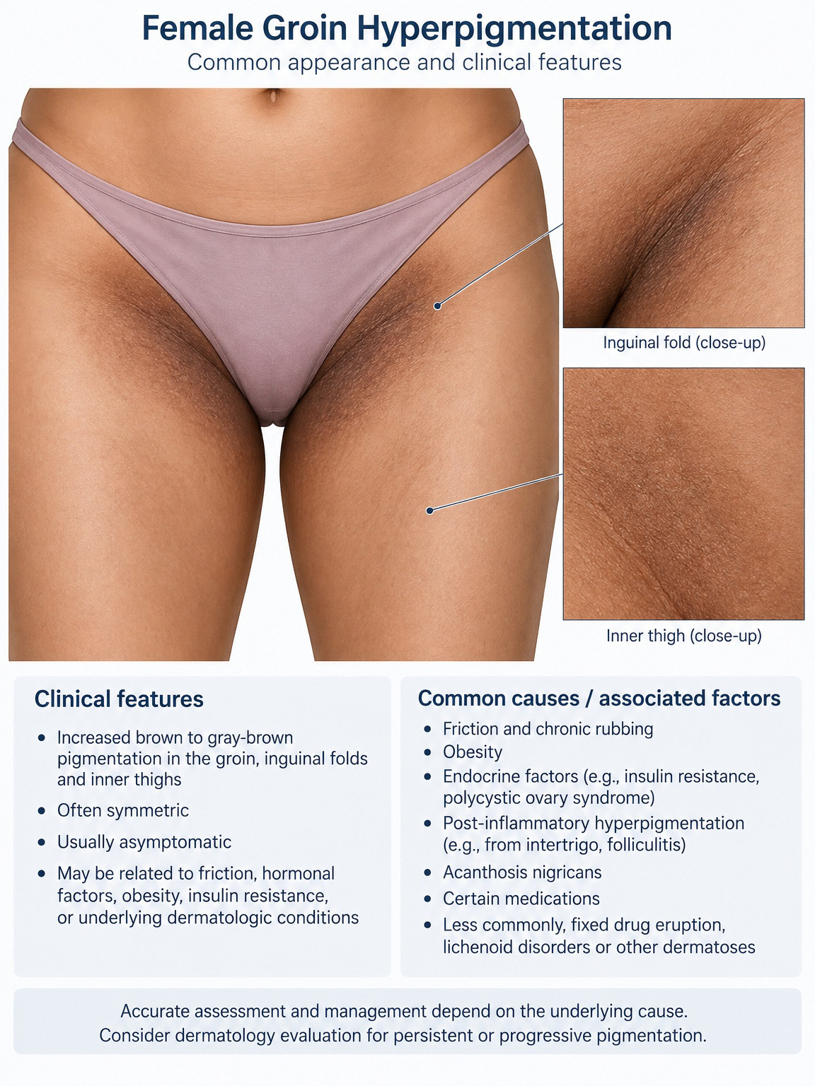
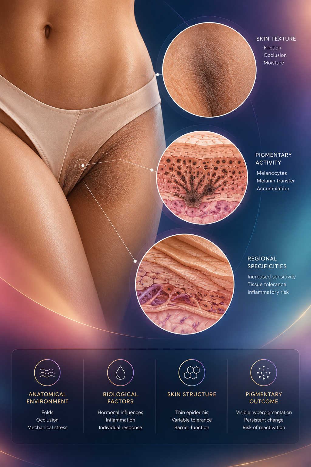
Clinical illustration showing intimate hyperpigmentation, regional skin biology and pigmentary activity
02
Clinical Topic Regional Skin Biology
Clinical Definition 02

Why Intimate Skin Is Different

Intimate hyperpigmentation describes increased visible pigmentation affecting external intimate and adjacent intertriginous skin. It should not be approached as ordinary facial or body pigmentation simply occurring in another location.

Clinical Distinction

Anatomical location changes the therapeutic context. Occlusion, friction, moisture, local sensitivity and the tendency toward inflammatory pigmentary change must all be considered before treatment intensity is selected.

01

Friction & Occlusion

Repeated mechanical contact and occlusive conditions may influence both pigmentation and treatment tolerance.

02

Variable Sensitivity

Tolerance is not uniform across intimate and adjacent cutaneous areas, making regional assessment essential.

03

Inflammatory Response

Irritation itself can perpetuate or intensify pigmentation, particularly in patients predisposed to pigmentary change.

04

Anatomical Precision

External cutaneous surfaces, folds and mucosal structures are not therapeutically interchangeable and must be distinguished clinically.

The first question is therefore not “How can this area be lightened?” but “What tissue are we treating, and why is it pigmented?”

Anatomical Mapping 03

One Clinical Term. Different Anatomical Areas.

“Intimate hyperpigmentation” may involve several external cutaneous regions. Each exists within a different anatomical environment and should therefore be identified precisely before treatment is considered.

The anatomical treatment field must be defined before the therapeutic pathway.
Essential Anatomical Boundary

External cutaneous surfaces and mucosal structures are anatomically distinct and must not be considered therapeutically interchangeable.

Identify Tissue First
Clinical Anatomy External Cutaneous Territories
Zoom
Medical illustration showing external anatomical areas that may present intimate hyperpigmentation
03
Anatomical Reference External Skin · Folds · Adjacent Areas
01
External Cutaneous Area Regional Precision

External Vulvar Skin

Pigmentation may involve external keratinized skin, particularly the labia majora and immediately adjacent cutaneous surfaces.

External Skin Variable Pigmentation Sensitivity
02
Intertriginous Area Fold Environment

Groin Folds

Groin pigmentation develops within an intertriginous environment where repeated contact, occlusion and moisture may influence the clinical presentation.

Friction Occlusion Moisture
03
Adjacent Cutaneous Area Extension Pattern

Inner Thighs

Pigmentation may extend beyond the folds onto adjacent thigh skin, where mechanical contact and previous inflammatory episodes may contribute to visible change.

Mechanical Contact PIH Distribution
04
External Cutaneous Area High Precision

Perianal Skin

External perianal pigmentation represents a distinct anatomical context in which the cutaneous treatment field must be defined with particular precision.

External Skin Boundaries Tolerance
Clinical Presentation

What Should Be Described Before Treatment?

Observe Before You Correct
01
Distribution Localized, diffuse or extending beyond the fold
02
Symmetry Symmetrical or predominantly unilateral
03
Colour Pattern Uniform, irregular or heterogeneous pigmentation
04
Skin Condition Intact, irritated or showing inflammatory change
03
From Anatomy to Strategy

Location defines the treatment field — not the protocol. Once the anatomical area has been identified, the next step is to determine the mechanism responsible for the pigmentation.

Anatomy → Mechanism
Anatomical Areas & Clinical Presentation
Pigmentary Drivers 04

What Drives Intimate Hyperpigmentation?

Visible pigmentation may be the final expression of several interacting biological and environmental influences. Identifying the dominant driver is more informative than describing colour alone.

The same visible pigmentation can arise from different clinical mechanisms.
01 Mechanical

Repetitive Friction

Recurrent rubbing or mechanical contact may maintain low-grade irritation and progressively reinforce visible pigmentation.

Repeated Contact → Irritative Signal → Pigmentary Change
02 Inflammatory

Post-Inflammatory Pigmentation

Previous irritation, dermatitis, follicular inflammation, shaving-related trauma or other inflammatory events may leave persistent pigmentary change after the visible inflammation has resolved.

Inflammation → Recovery → Residual Pigment
Clinical Expression Persistent Pigmentary Signal

Often multifactorial rather than driven by one isolated cause.

Mechanism Before Treatment

Determine what continues to stimulate or maintain the pigmentation before deciding how aggressively it should be corrected.

03 Hormonal

Hormonal Influence

Hormonal context may influence regional pigmentation and contribute to variation over time, particularly when other local pigmentary stimuli are also present.

Hormonal Context → Regional Influence → Visible Change
04 Individual

Constitutional Pigmentary Tendency

Baseline pigmentation and individual reactivity influence both the clinical appearance and the tendency to develop additional pigmentation after irritation.

Baseline Biology → Individual Response → Variable Expression
05
Perpetuating Factor

Repeated External Irritation

Hair removal, aggressive cleansing, unsuitable topical products and repeated cosmetic manipulation may become continuing sources of irritation. In such cases, treatment without correcting the trigger may reproduce the same pigmentary environment.

External Trigger → Repeated Irritation → Recurrence
Clinical Reality

Mechanisms Frequently Overlap

Friction + Inflammation + Individual Reactivity = Persistent Pigmentation

A multifactorial presentation should not be reduced to a single presumed cause. The relative importance of each driver must be established clinically.

From Mechanism to Assessment

Once the likely drivers have been identified, the next step is to determine whether the patient and the affected tissue are appropriate for treatment.

Mechanism → Assessment
Pre-Treatment Decision 05

Clinical Assessment & Treatment Eligibility

Identifying pigmentation is not sufficient to justify immediate treatment. The clinician must first determine whether the tissue is suitable for intervention, whether relevant risk factors are controlled and whether the clinical presentation requires further evaluation.

Assessment determines whether to treat, when to treat and when not to treat.
Clinical Gate Four decisions before treatment
Assess → Exclude → Stratify → Decide
01
First Gate

Assess

Observe

Establish the current condition of the treatment field before considering any corrective intervention.

✓

Skin integrity Intact surface without clinically significant irritation or disruption.

✓

Current clinical activity Determine whether erythema, inflammation, discomfort or other active changes are present.

✓

Clinical history Document onset, evolution, previous procedures and prior responses to topical or procedural treatment.

02
Safety Gate

Exclude

Verify

Pigmentation should not automatically be interpreted as an indication for a cosmetic or corrective procedure.

!

Active inflammatory disease Defer treatment when clinically significant inflammation is present.

!

Infection or suspicious lesion Findings requiring diagnosis or medical management take priority over pigment correction.

!

Uncertain diagnosis Atypical, evolving or unexplained pigmentation should be clarified before elective treatment.

03
Risk Gate

Stratify

Estimate

Estimate individual susceptibility before choosing treatment intensity or therapeutic pathway.

+

Phototype Record phototype as one component of the overall pigmentary risk assessment.

+

History of PIH Previous post-inflammatory pigmentation may indicate greater susceptibility to pigmentary rebound.

+

Previous tolerance Consider the response to earlier procedures, irritation and recovery rather than treatment history alone.

04
Decision Gate

Decide

Select

Integrate the clinical findings before moving from assessment to treatment planning.

→

Treatment readiness Determine whether the tissue is clinically suitable for intervention now.

→

Need for stabilization Correct modifiable local conditions before attempting pigment correction when necessary.

→

Need for referral Do not proceed when diagnosis, pathology or clinical findings require investigation beyond the intended procedure.

Patient-Specific Context

Clinical Eligibility Is More Than Skin Examination

Treatment planning should also account for circumstances that may alter tolerance, recovery, adherence or the interpretation of the expected result.

01
Recent Procedures Hair removal, exfoliation or other recent local procedures.
02
Current Topicals Products capable of irritating or altering local tolerance.
03
Recovery Conditions Ability to minimize avoidable irritation during recovery.
04
Patient Expectations Goals should be clinically realistic and compatible with gradual pigmentary management.
Treatment Eligibility

Three Possible Clinical Decisions

Assessment → Action
✓
Eligible

Proceed

The treatment field is clinically suitable and no identified finding requires prior stabilization or further investigation.

≈
Temporarily Deferred

Stabilize First

Modifiable irritation, inflammation or another temporary condition should be controlled before reassessment.

!
Further Evaluation

Do Not Treat & Refer

Uncertain diagnosis, suspicious findings or relevant pathology requires appropriate medical evaluation before elective pigment treatment.

05
Eligibility Before Strategy

A treatment pathway begins only after eligibility has been established. The next step is to understand which pigmentary compartments should be targeted and regulated.

Eligibility → Pigment Model
Metabolic Pigmentation Science 06

Tenenbaum–Tiziani Metabolic Pigmentation Model

Visible pigmentation represents more than the presence of melanin. It reflects a dynamic biological sequence involving pigment formation, cellular transfer and the progressive handling of pigment already present within the epidermal system.

Effective pigment management can therefore be considered across several interconnected metabolic compartments.
TT
Core Concept

Pigmentation Is a Dynamic System

The therapeutic objective is not limited to removing visible pigment. A metabolic strategy considers how pigment is produced, how it is transported and how existing pigment is progressively degraded and cleared.

Mechanism → Compartment → Strategy
01
Compartment One

Melanin Production

The first compartment concerns the biological activity responsible for pigment formation. Excessive or persistent melanogenic activity can continue to replenish visible pigmentation even when existing surface pigment is reduced.

Therapeutic Focus Regulate excessive pigment formation
02
Compartment Two

Melanin Transport

Pigment must also be considered in terms of its transfer and distribution within the epidermal system. Regulation at this level addresses the movement of melanin rather than its synthesis alone.

Therapeutic Focus Modulate pigment transfer and distribution
03
Compartment Three

Degradation & Clearance

Existing pigment represents a third therapeutic dimension. Progressive epidermal processing and elimination influence how long previously formed pigment remains clinically visible.

Therapeutic Focus Facilitate progressive handling of existing pigment
Transversal Modulator

Inflammatory Signalling

Inflammatory signalling is not presented here as a fourth melanin compartment. It acts across the pigmentary system and may modify melanogenic activity, tissue response and the persistence of pigmentary change.

Production Transport Clearance
Conceptual Difference

Visible Pigment vs. Pigment Dynamics

These two perspectives should not be confused. One describes what is visible at a given moment; the other considers the biological processes that continually determine that visible result.

A Static Perspective

Visible Pigment

Focuses primarily on pigment already clinically apparent within the treatment field.

What is visible now
VS
B Metabolic Perspective

Pigment Dynamics

Considers the continuing balance between pigment formation, transfer and progressive clearance.

What determines pigmentation over time
TT
Tenenbaum–Tiziani Principle

Three Compartments. One Pigmentary System.

Production + Transport + Degradation / Clearance = Pigmentary Balance

The relative importance of each compartment may differ between patients and clinical presentations. This provides a framework for selecting a therapeutic strategy rather than assuming that every pigmentation problem requires the same form of correction.

From Model to Treatment

Once the pigmentary system is understood, treatment can follow different therapeutic pathways according to the clinical objective and the required degree of correction or regulation.

Metabolic Model → Two Pathways
Therapeutic Strategy 07

Two Therapeutic Pathways

Once treatment eligibility and the pigmentary model have been established, the therapeutic objective can follow two distinct but potentially complementary pathways: controlled correction of visible pigmentation or progressive metabolic regulation.

The choice is strategic: correct what is visible, regulate what sustains pigmentation, or combine both approaches sequentially.
A B
Therapeutic Principle

Different Objectives. Different Biological Logic.

A corrective pathway primarily addresses clinically visible pigment through a controlled procedural endpoint. A metabolic pathway addresses pigmentary dynamics progressively over time.

Select the Objective First
A
Corrective Pathway

Controlled Correction

Frosting-Based

A more directly corrective strategy directed toward visible pigmentation, using a deliberately controlled clinical endpoint within an appropriately selected treatment field.

01 Visible Pigment Defined corrective target
→
02 Controlled Action Deliberate treatment intensity
→
03 Clinical Endpoint Observable procedural response
✦
Key Concept Frosting is an endpoint — not a treatment objective by itself.

Its significance depends on the selected agent, anatomical site, tissue tolerance and intended depth of controlled correction.

OR AND
B
Regulatory Pathway

Metabolic Regulation

Progressive

A progressive strategy directed toward the biological dynamics that determine pigmentation rather than toward visible pigment alone.

01 Pigment Dynamics Metabolic framework
→
02 Regulation Progressive modulation
→
03 Pigmentary Balance Longitudinal objective
∞
Key Concept Regulation addresses pigment dynamics rather than pigment visibility alone.

It may target one or more pigmentary compartments described in the Tenenbaum–Tiziani metabolic model.

Clinical Integration

Not Necessarily Either / Or

The two pathways describe different therapeutic objectives, but they are not necessarily mutually exclusive. In selected cases, controlled correction may be followed by progressive regulation of the pigmentary environment.

01 Initial Objective Correction
→
02 Biological Objective Regulation
→
03 Long-Term Objective Maintenance
!

This sequence represents a therapeutic concept, not a universal protocol. The appropriate pathway depends on the clinical assessment, treatment field and intended objective.

Strategic Comparison

Two Pathways, Two Primary Objectives

Primary Focus
Visible established pigmentation
Pigmentary dynamics
Approach
Controlled corrective intervention
Progressive biological regulation
Endpoint Logic
Observable controlled response
Progressive change over time
Strategic Role
Correction
Regulation
Controlled Correction Metabolic Regulation
A B
From Strategy to Selection

Once the therapeutic pathway has been defined, the next question is which product and protocol best match that objective.

Pathway → Product & Protocol
Clinical Selection Framework 08

Product & Protocol Selection

Product selection follows the therapeutic objective. Corrective acids, post-acid transition products and metabolic regulators do not occupy the same position within the treatment sequence.

Select the pathway first, then place each product in its correct clinical sequence.
Essential Sequence

When a Corrective Acid Is Used

The order of application is fundamental. Peeling de Luxe Plus occupies the transition between the corrective acid step and subsequent metabolic products.

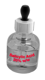
01 Corrective Step TCA / Salicylic-Type Acid

Controlled acid application according to the selected corrective objective.

→
02 Mandatory Sequence Position Peeling de Luxe Plus

Applied after the acid step and before subsequent metabolic products.

→
03 Metabolic Step Metabolic Products

Selected according to the intended pigment-regulating objective.

!

Sequence rule: Peeling de Luxe Plus is positioned after an acid such as TCA or salicylic acid and before the application of other metabolic products.

01
Corrective Family

Controlled Acid Correction

Used when the selected therapeutic pathway includes a direct corrective step against established visible pigmentation.

TCA corrective peeling product
Corrective Acid Step

TCA-Based Correction

A controlled corrective option when a frosting-based pathway has been selected and the anatomical treatment field is considered suitable.

Position Before Peeling de Luxe Plus
Salicylic acid corrective peeling product
Corrective Acid Step

Salicylic-Type Correction

An alternative corrective acid approach when clinically appropriate within the selected treatment strategy.

Position Before Peeling de Luxe Plus
02
Post-Acid Transition

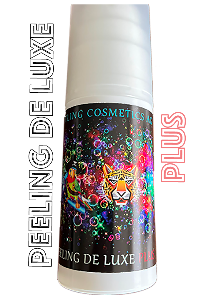
Peeling de Luxe Plus

A specific intermediate step within the corrective sequence, positioned after the acid application and before subsequent metabolic products.

Sequence Position Acid → Peeling de Luxe Plus → Metabolic
Peeling de Luxe Plus post-acid transition product
Peeling de Luxe Plus · Post-Acid Transition
Critical Sequence Step

After the Acid. Before Metabolic Products.

Peeling de Luxe Plus is not positioned as an isolated first-line acid step in this sequence. Its role follows the corrective acid application and precedes the products selected for subsequent metabolic management.

01

Post-Acid Position Used after the selected corrective acid step.

02

Frosting-Control Role Marks the controlled transition following the corrective acid phase.

03

Pre-Metabolic Position Applied before subsequent metabolic products.

!

Do not reverse the sequence: metabolic products belong after Peeling de Luxe Plus when the protocol includes a preceding corrective acid.

03
Metabolic Family

Progressive Pigment Regulation

Metabolic products are selected according to the pigmentary objective and their intended role within the regulatory strategy.

Clarté de Lune metabolic pigmentation treatment
Metabolic Pigment Regulation

Clarté de Lune

Positioned within the metabolic strategy for regulation of melanocyte activity and melanin-related pigmentation processes.

Clinical Role Melanocyte / Melanin Regulation
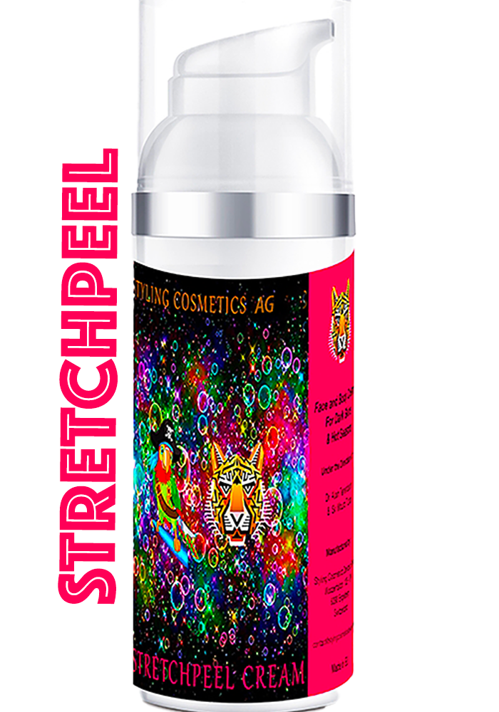
StretchPeel depigmenting metabolic treatment
Depigmenting Metabolic Protection

StretchPeel

A depigmenting component of the metabolic strategy, combining progressive pigment management with biological photoprotection against UVA- and UVB-related environmental stimulation.

Clinical Role Depigmentation + Biological Photoprotection
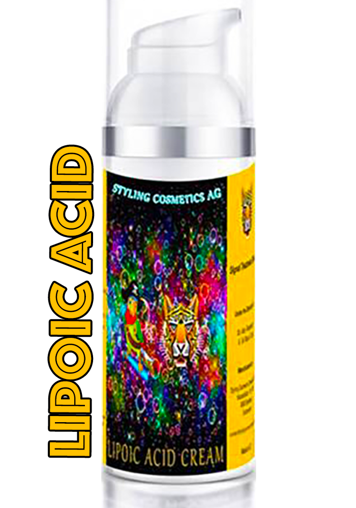
Lipoic Acid Cream metabolic treatment
Metabolic Penetration Support

Lipoic Acid Cream

Used within a metabolic strategy where enhanced penetration of associated active components forms part of the intended treatment logic.

Clinical Role Penetration Facilitator
Clinical Selection Logic

Product Choice Follows the Pathway

The same product sequence is not required for every patient. Selection depends on whether the objective is direct correction, progressive metabolic regulation or a planned combination of both.

A Corrective Pathway

Acid-Based Correction

Corrective Acid → Peeling de Luxe Plus

Selected when controlled correction of established visible pigmentation is the principal initial objective.

B Metabolic Pathway

Progressive Regulation

Metabolic Selection → Progressive Regulation

Selected when the therapeutic objective is progressive regulation rather than an initial frosting-based corrective step.

A+B Combined Strategy

Correction + Regulation

Acid → Peeling de Luxe Plus → Metabolic

Used when controlled correction is deliberately integrated with subsequent metabolic management.

01 02 03
Sequence Matters

Acid First. Transition Second. Metabolic Products After.

When the selected protocol begins with TCA, salicylic acid or another appropriate corrective acid, Peeling de Luxe Plus follows that acid step. Other metabolic products are applied only after this transition step within the intended protocol.

01 02 03
From Selection to Strategy

Product selection defines the therapeutic tools. The next step is to organize them longitudinally through correction, stabilization and maintenance.

Product Selection → Treatment Strategy
Longitudinal Treatment Strategy 09

From Correction to Pigmentary Stability

Successful pigment management is not defined by the initial lightening response alone. Treatment should evolve through successive clinical phases as visible pigmentation decreases and biological stability improves.

The objective is to move from active correction toward durable control with progressively less therapeutic intensity.
01 02 03
Core Treatment Principle

Treatment Changes as the Pigmentary State Changes.

The strategy should not remain therapeutically static. The objective of the initial phase differs from that of stabilization, and long-term maintenance requires a different level of intervention again.

Dynamic Strategy
Therapeutic Evolution

Three Phases. One Continuous Strategy.

Each phase has a different clinical objective, but all three belong to the same longitudinal management plan.

01
Initial Phase

Correction

Reduce

Address the established visible pigmentary burden with the degree of intervention justified by the selected clinical pathway and tissue tolerance.

Primary Objective Controlled improvement of visible pigmentation

Begin from the treatment objective already established during assessment and pathway selection.

Correct without exceeding the response appropriate for the anatomical treatment field.

Evaluate the clinical response before deciding whether further corrective intensity is justified.

Transition When The corrective objective has been sufficiently achieved to shift the priority from reduction to stabilization.
Shift →
02
Consolidation Phase

Stabilization

Consolidate

After visible improvement, the therapeutic priority moves toward consolidation of the response and reduction of conditions that could rapidly recreate the previous pigmentary state.

Primary Objective Prevent early pigmentary reactivation

Allow the treated tissue to progress toward a more stable clinical state.

Continue regulation where the pigmentary environment remains biologically active.

Avoid unnecessary repetition of corrective intensity when consolidation is the principal objective.

Transition When Improvement remains clinically stable and active correction is no longer the dominant requirement.
Shift →
03
Long-Term Phase

Maintenance

Preserve

Once a satisfactory and stable pigmentary state has been obtained, management shifts toward preservation rather than repeated active correction.

Primary Objective Preserve long-term pigmentary balance

Maintain the improvement with the lowest appropriate therapeutic burden.

Continue control of relevant local or environmental factors that may favour recurrence.

Reassess if pigmentation begins to change rather than automatically restarting intensive correction.

Long-Term Goal Stable improvement with progressively less need for active intervention.
Therapeutic Balance

As Stability Increases, Treatment Intensity Should Decrease.

The longitudinal strategy is not based on maintaining the same intervention indefinitely. Therapeutic intensity should adapt to the changing clinical state.

Therapeutic Intensity
Higher → Lower
Pigmentary Stability
Lower → Higher
Correction Stabilization Maintenance
Clinical Evolution

The Endpoint of One Phase Becomes the Starting Point of the Next.

01 Correction Visible Pigmentation ↓

Controlled improvement

02 Stabilization Improved Pigmentary State ↓

Consolidated response

03 Maintenance Stable Pigmentary State ↓

Long-term preservation

TT
Longitudinal Principle

Successful Treatment Does Not End When Pigmentation Becomes Lighter.

Initial correction changes what is clinically visible. Stabilization determines whether that improvement can be consolidated. Maintenance determines whether the resulting pigmentary balance can be preserved over time.

Clinical Adaptation

The Timeline Is Individual.

These phases describe therapeutic objectives rather than fixed calendar periods. Progression depends on clinical response, tissue recovery and pigmentary stability.

01 Response-Driven

Move between phases according to the observed clinical response rather than a predetermined date alone.

02 Tissue-Driven

Tissue tolerance and recovery remain essential when deciding whether further intervention is appropriate.

03 Stability-Driven

Long-term management should reflect the degree of pigmentary stability already achieved.

01 02 03
From Strategy to Procedure

Once the therapeutic phase and objective have been defined, the next step is to organize the clinical procedure and treatment sequence.

Strategy → Procedure
Clinical Procedure · 10

Clinical Procedure & Treatment Sequence

Intimate pigmentation treatment requires a controlled, anatomically defined and response-guided procedure. The clinical sequence depends on the therapeutic pathway selected, while tissue tolerance and the observed response determine how the session progresses.

Procedural Principle

The procedure is not defined by the pursuit of maximal visible reaction. Each step should remain proportional to the anatomical treatment field, the selected corrective or regulatory pathway, and the response observed during treatment.

Session Architecture

A Controlled Clinical Sequence

01

Prepare

Define and prepare the external cutaneous treatment field according to the clinical assessment.

02

Select

Confirm whether the session follows a corrective acid-based pathway or a primarily regulatory pathway.

03

Apply

Perform the selected intervention within the previously defined external cutaneous treatment field.

04

Observe

Read the tissue response continuously and interpret visible endpoints in their clinical context.

05

Complete

Complete the session according to the selected pathway, tissue response and regulatory objective.

Clinical Decision Pathway

One Assessment — Two Procedural Routes

Not every treatment session requires the same procedural route. The pathway follows the clinical objective established during assessment.

Starting Point External Cutaneous Treatment Field Defined
Clinical Decision Is a Corrective Acid-Based Step Indicated?
Yes · Corrective Route

Controlled Correction

When an acid-based corrective step has been selected, application is controlled and the tissue response determines progression through the sequence.

1 Corrective Acid TCA or salicylic-type corrective step when clinically selected.
2 Observe Clinical Endpoint Read the visible tissue response without treating frosting as a universal objective.
3 Peeling de Luxe Plus Positioned after the acid step and before other metabolic products.
4 Metabolic Step Continue with the selected metabolic products according to the regulatory strategy.
No · Regulatory Route

Metabolic Regulation

When a corrective acid step is not selected, the session can proceed through the appropriate metabolic strategy without artificially reproducing the corrective sequence.

1 Confirm Regulatory Objective Match the intervention to the pigmentation mechanism and current treatment phase.
2 Select Metabolic Approach Use the product family appropriate to the intended pigmentary regulation.
3 Respect Tissue Tolerance Keep the intervention proportional to the anatomical field and observed response.
4 Complete the Session Conclude according to the selected regulatory strategy and immediate clinical response.
Pathway Convergence Both routes return to clinical observation and longitudinal regulation.

The procedural route may differ, but treatment remains guided by anatomy, tissue response and the evolving pigmentary state.

Clinical Endpoint Recognition

Read the Tissue Response

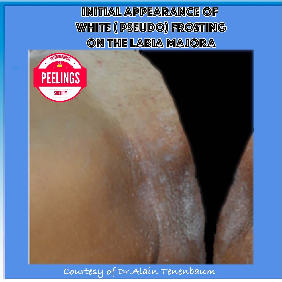
During a selected corrective procedure, the initial appearance of white or pseudo-frosting provides clinical information about the local response.

It should be interpreted dynamically and in relation to the anatomical site, baseline pigmentation and tissue tolerance. A more pronounced visible reaction does not automatically represent a better therapeutic result.

Observe the onset and distribution of the visible response.
Interpret the appearance together with tissue tolerance.
Do not equate greater visible frosting with greater efficacy.
Initial Clinical Endpoint + Zoom
Initial appearance of white pseudo-frosting on external labia majora skin during an intimate peeling procedure
Initial white / pseudo-frosting appearance. Clinical observation of an early visible response on external labia majora skin during a selected corrective procedure.
Clinical Endpoint Principle

Observe the Endpoint — Do Not Chase It

Frosting or pseudo-frosting, when present, is an observable clinical sign rather than a universal therapeutic objective. Its appearance, distribution and evolution must be interpreted together with the anatomical site and the overall tissue response.

Corrective Pathway · Fixed Order

The Post-Acid Sequence Matters

STEP 01 Corrective Acid TCA or salicylic-type corrective step when selected
›
STEP 02 Clinical Endpoint Observe and interpret the tissue response
›
STEP 03 Peeling de Luxe Plus Post-acid transition before other metabolic products
›
STEP 04 Metabolic Step Continue according to the selected regulatory strategy

When an acid-based corrective pathway is used, the order is preserved: corrective acid first, followed by clinical endpoint assessment, then Peeling de Luxe Plus, and only afterwards the selected metabolic products.

!
Anatomical Safety Boundary

External Cutaneous Tissue ≠ Mucosal Tissue

The treatment field described here concerns external cutaneous intimate areas. Mucosal structures and external skin are not therapeutically interchangeable and must not be approached as if they shared the same treatment tolerance.

Next · Expected Evolution
From immediate response to the days and weeks that follow.
Clinical Evolution · 11

Expected Evolution & Clinical Endpoints

The immediate appearance of treated skin represents only one moment in the clinical process. A visible endpoint during treatment must be distinguished from the biological evolution that follows. Assessment therefore continues beyond the treatment session.

Evolution Principle

Clinical interpretation should follow the complete sequence: immediate tissue response, early post-treatment evolution, progressive recovery and subsequent pigmentary reassessment. No isolated visual stage should be interpreted as the final treatment result.

Clinical Time Course

From Immediate Response to Reassessment

D0

Immediate Endpoint

The initial visible tissue response is documented and interpreted within the context of the procedure performed.

EARLY

Post-Treatment Evolution

The treated field evolves after the session. Its appearance should not be judged solely against the immediate endpoint.

RECOVERY

Progressive Resolution

As the local response settles, the underlying pigmentary pattern becomes progressively more suitable for reassessment.

REVIEW

Pigmentary Reassessment

The next clinical decision is based on the evolved tissue state rather than on the appearance seen during treatment.

Serial Clinical Observation

Evolution Is a Sequence, Not a Snapshot

Serial photography is particularly useful because the appearance observed immediately after treatment does not represent the final pigmentary state.

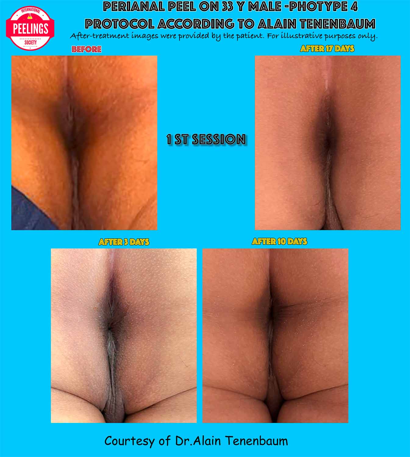
This documented first-session example follows the treated perianal area through successive observations and demonstrates why clinical interpretation must extend beyond the day of treatment.

The relevant question is therefore not simply “What does the skin look like now?” but “How is the treated field evolving over time?”

First Session · Serial Evolution + Zoom
Serial clinical evolution of a perianal peeling treatment following the first session
Serial post-treatment documentation. Clinical evolution following the first perianal peeling session, illustrating successive stages rather than an isolated immediate endpoint.
Clinical Reading

Four Questions During Follow-Up

01

Is the Response Settling?

Follow the local tissue response rather than interpreting the immediate post-treatment appearance as permanent.

02

Is the Field Recovering?

Reassessment should consider how the treated cutaneous field progresses through its post-treatment evolution.

03

What Pigment Remains?

Once the transient treatment response has evolved, residual pigmentation can be interpreted more meaningfully.

04

What Comes Next?

The evolved clinical state determines whether the subsequent strategy requires further correction, regulation or observation.

Second Session · Pseudo-Frosting + Zoom
Pseudo-frosting observed during a second perianal peeling session
Pseudo-frosting during a subsequent session. The visible treatment endpoint provides immediate clinical information, but should not be confused with the eventual pigmentary outcome.
Endpoint Interpretation

Endpoint ≠ Outcome

Pseudo-frosting is an immediate clinical observation. It documents the response occurring during a selected corrective procedure at that particular moment.

The longer-term clinical outcome is evaluated later, after the treated field has evolved and the transient procedural response no longer dominates its appearance.

This distinction prevents a visible intra-procedural sign from being mistaken for the therapeutic result itself.

Essential Distinction

Clinical Endpoint Is Not Clinical Outcome

During Treatment

Clinical Endpoint

A visible tissue response observed during the procedure, including frosting or pseudo-frosting when present.

→
After Evolution

Clinical Outcome

The later condition of the treated field after the immediate procedural response has evolved and pigmentation can be reassessed.

Reassessment Principle

Treat the Evolved Clinical State — Not the Previous Session

Each subsequent decision should be based on the tissue and pigmentation actually present at reassessment. Previous treatment intensity, previous frosting and previous appearance do not replace examination of the current clinical state.

Next · Phototypes & Safety
Clinical evolution must always be interpreted through individual PIH risk.
Risk Stratification · 12

Phototype & PIH Risk Stratification

Pigmentary reactivity is not determined by phototype alone. In intimate areas, the risk of post-inflammatory hyperpigmentation must be interpreted as a combined clinical profile involving baseline pigmentation, previous inflammatory responses, local irritation and the intensity of the proposed intervention.

Risk-Stratification Principle

Fitzpatrick phototype contributes useful information about cutaneous pigmentary behaviour, but it should not be used as an isolated prediction of PIH. Individual history, inflammation, friction, tissue condition and treatment-related factors can substantially modify the clinical risk.

PIH Risk Stratification Map

Risk Emerges From Multiple Converging Factors

The objective is not to assign risk from skin colour alone, but to integrate the factors that can amplify pigmentary reactivity after inflammation.

FACTOR 01

Phototype

Baseline cutaneous pigmentation and pigmentary response provide one component of the overall risk profile.

FACTOR 02

Previous PIH

A personal history of hyperpigmentation following inflammation or procedures is clinically relevant.

FACTOR 03

Active Inflammation

Ongoing inflammatory activity can increase melanogenic signalling and alter the response to treatment.

FACTOR 04

Friction & Trauma

Repetitive mechanical irritation may maintain both inflammation and the pigmentary stimulus.

FACTOR 05

Current Skin State

Barrier condition, irritation and local tissue tolerance modify the suitability of a corrective intervention.

FACTOR 06

Proposed Intervention

The expected inflammatory burden of the selected procedure must be considered within the individual pigmentary profile.

Integrated Clinical Profile

Individual PIH Susceptibility

No single factor defines the patient. Risk is interpreted from the interaction between pigmentary biology, inflammatory history, local anatomy and the proposed treatment.

Therapeutic Consequence Greater PIH Concern → Greater Need to Limit Unnecessary Inflammation

Risk stratification modifies the balance between corrective intensity, metabolic regulation and the timing of reassessment.

Phototype in Context

Phototype Is a Modifier — Not the Whole Risk Profile

Fitzpatrick classification remains clinically useful, but its original function and an individual's tendency to develop post-inflammatory pigmentation should not be treated as interchangeable concepts.

Two patients with a similar phototype may have very different histories of PIH, friction, inflammation and tolerance to previous procedures.

For this reason, phototype should contribute to clinical stratification without replacing the individual history and examination.

Fitzpatrick I–VI

One Variable Within a Larger Clinical Model

I Phototype
II Phototype
III Phototype
IV Phototype
V Phototype
VI Phototype
Do not convert this scale into an automatic PIH score. Phototype must be interpreted together with the patient's actual inflammatory and pigmentary history.
Beyond Phototype

Factors That Can Shift the Risk Profile

01

Previous PIH

Previous pigmentation after inflammation, trauma or procedures provides direct information about individual pigmentary reactivity.

02

Recurrent Friction

Persistent rubbing or mechanical irritation may continue to generate an inflammatory stimulus even after treatment.

03

Inflammatory Activity

Active irritation changes the biological context in which a pigment-corrective intervention would occur.

04

Recent Trauma

Recent mechanical, procedural or other local trauma can modify both tissue tolerance and pigmentary behaviour.

05

Barrier Condition

The current state of the external cutaneous barrier forms part of the decision to correct, regulate or defer.

06

Treatment Intensity

The potential inflammatory burden of the proposed intervention must remain proportional to the individual risk profile.

Clinical Stratification

Risk Changes the Therapeutic Balance

Lower PIH Concern

Standard Clinical Caution

When the combined profile is favourable, treatment can follow the selected pathway while remaining guided by anatomy and observed tissue response.

Intermediate PIH Concern

Increase Procedural Restraint

As pigmentary concern increases, unnecessary inflammatory burden becomes progressively less acceptable and reassessment gains greater importance.

Higher PIH Concern

Prioritize Pigmentary Control

A more reactive clinical profile shifts the therapeutic balance toward conservative correction, metabolic regulation and careful longitudinal reassessment.

Therapeutic Logic

As PIH Concern Increases, Strategy Must Adapt

Higher Pigmentary Reactivity Integrated clinical risk profile
›
Greater Consequence of Inflammation Avoid unnecessary inflammatory burden
›
More Conservative Correction Treatment intensity follows tolerance
›
Greater Regulatory Emphasis Metabolic strategy and reassessment
Clinical Rule

Stratify the Patient — Not the Phototype Alone

Phototype contributes to risk assessment, but treatment decisions should follow the complete clinical profile. The most relevant question is not simply how pigmented the skin is, but how that individual's skin has responded to inflammation, trauma and previous interventions — and how much inflammatory burden the proposed treatment may create.

Next · Contraindications
Risk modifies treatment — contraindications determine when not to treat.
Anatomical Safety · 13

Contraindications & Areas Not to Treat

Intimate pigmentation may extend across adjacent anatomical structures that are not biologically equivalent treatment surfaces. The first safety decision is therefore not which product to apply, but precisely where the external cutaneous treatment field ends.

Anatomical Safety Principle

External anatomical location does not automatically mean external cutaneous tissue. Treatment must remain confined to an anatomically identified external skin field. Transitional, mucosal and internal epithelial structures must not be considered interchangeable with keratinized external skin.

Adjacent Structures — Different Treatment Status

The external vulvar region contains different tissue types. Anatomical proximity must never be interpreted as therapeutic equivalence.

Potential Cutaneous Field

External Labia Majora

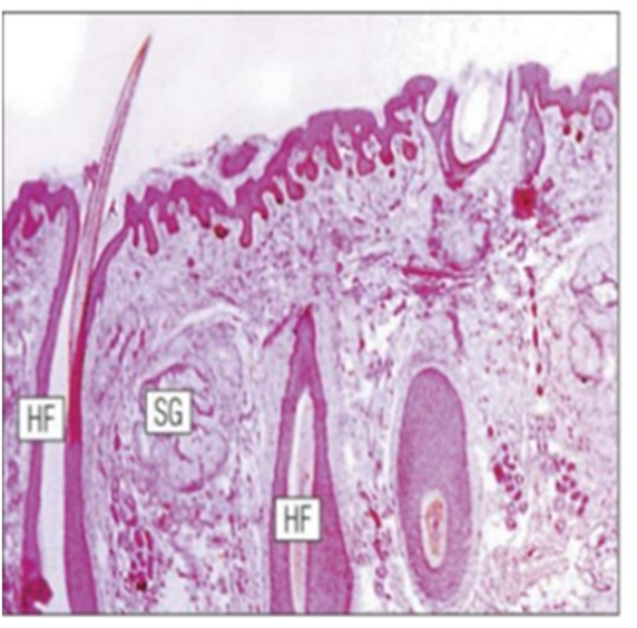
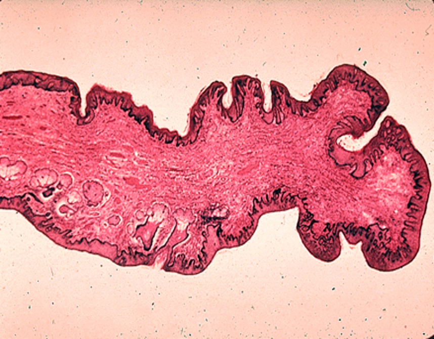
Histological appearance of external labia majora skin showing cutaneous structures and hair follicles

The external, hair-bearing surface of the labia majora presents cutaneous characteristics including hair follicles and sebaceous structures. When clinically appropriate, this external skin may form part of a precisely delimited treatment field.

Do Not Treat

Labia Minora

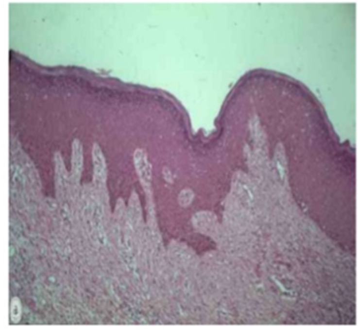
Histological appearance of labia minora tissue

The labia minora must not be treated as though they were simply an extension of external hair-bearing labia majora skin. For the protocols described here, they remain outside the external cutaneous peeling field.

Do Not Treat

Vulvar Vestibule

Histological appearance of vulvar vestibular tissue

Vestibular and internal vulvar surfaces are not equivalent to external cutaneous tissue. They constitute an anatomical exclusion zone for the external pigmentation protocols described on this page.

Practical Anatomical Landmark

Hair-bearing skin can provide a useful landmark for identifying the external cutaneous field, but hair presence or absence alone does not define tissue type. The treatment boundary must ultimately follow anatomy and tissue characteristics, not hair distribution alone.

External Skin Hair-Bearing Labia Majora
›
Precision Boundary Medial Transition
›
Exclusion Zone Labia Minora
›
Exclusion Zone Vestibule / Internal Surfaces
Perianal Anatomical Stop-Line

The External Treatment Field Has a Defined Limit

Perianal skin, the external anal margin and internal anal structures must be anatomically distinguished before any treatment is considered.

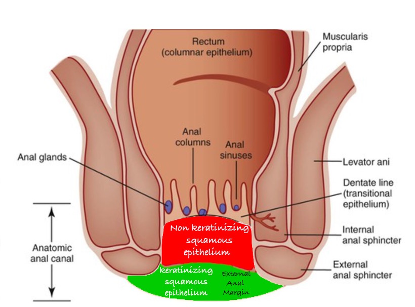
External Anal Margin · Transition Anatomy + Zoom
Anatomical diagram showing external anal margin, keratinized external squamous epithelium, transitional anal epithelium and rectal structures
Anatomical transition at the anal margin. External keratinized cutaneous tissue must be distinguished from the transitional and internal epithelial structures of the anal canal.

Anatomy Defines the Stop Line

The treatment field progresses only across appropriately selected external cutaneous tissue. Movement toward the anal canal reaches an anatomical boundary beyond which the external pigmentation protocol must not continue.

Perianal Skin Potential external cutaneous treatment field.
External Anal Margin High-precision anatomical boundary requiring exact delimitation.
Anatomical Stop Line
Internal Anoderm / Anal Canal Outside the external cutaneous pigmentation treatment field.
Rectal Mucosa Internal anatomy — never an external peeling treatment field.
Anatomical Exclusion Zones

Areas Not to Treat

Labia Minora

Excluded from the external cutaneous peeling field described in this clinical approach.

Vulvar Vestibule

Internal vestibular surfaces must not be treated as external keratinized skin.

Vaginal Introitus & Vaginal Mucosa

Vaginal mucosal structures are outside the treatment field of the external pigmentation protocols described here.

Clitoral & Internal Preputial Surfaces

Clitoral structures and internal surfaces of the clitoral hood are not treatment fields for these external peeling protocols.

Periurethral / Urethral Region

Urethral and immediately periurethral internal structures must remain outside the external treatment field.

Internal Anal Canal & Rectal Mucosa

Treatment of external perianal pigmentation must not extend into the internal anal canal or rectal mucosa.

Clinical Contraindications

When the Anatomical Field Should Not Be Treated

Even anatomically appropriate external skin may be unsuitable for treatment when the local clinical condition is not compatible with a controlled procedure.

Active Inflammation

Active inflammatory disease or significant ongoing irritation requires evaluation before treatment.

Infection or Suspected Infection

Active or suspected local infection is incompatible with elective pigment-corrective treatment.

Erosion / Ulceration

Eroded, ulcerated or otherwise non-intact tissue should not receive the external peeling procedure.

Significant Barrier Disruption

A compromised external cutaneous barrier requires recovery and reassessment before treatment is considered.

Recent Local Trauma or Procedure

Recently traumatized or procedurally altered tissue should not be treated until clinically appropriate for reassessment.

Undiagnosed Lesion

An unexplained lesion or atypical pigmentation requires diagnostic clarification rather than empirical peeling.

Final Anatomical Rule

Never Extend the Protocol Simply Because Pigmentation Continues

Pigmentation may cross from external skin toward transitional or mucosal structures, but the treatment field must not follow it indiscriminately. Anatomy — not the visible extent of pigmentation — defines the treatment boundary.

Detailed Clinical Protocol

Perianal Treatment Protocol

For detailed regional anatomy, external anal margin delimitation and the complete clinical approach to the perianal area, consult the dedicated protocol.

View Perianal Protocol →
Next · Clinical Cases
From anatomical safety boundaries to documented clinical outcomes.
Clinical Documentation · 14

Clinical Cases / Before & After

Clinical photography provides a longitudinal view of treatment response. The case below documents baseline pigmentation, the recorded treatment course and long-term follow-up within the context of an individual clinical outcome.

Clinical Documentation Principle

A before-and-after comparison should be interpreted together with the anatomical area, baseline pigmentation, treatment course and follow-up interval. A final photograph alone does not describe the biological evolution that occurred between the two observations.

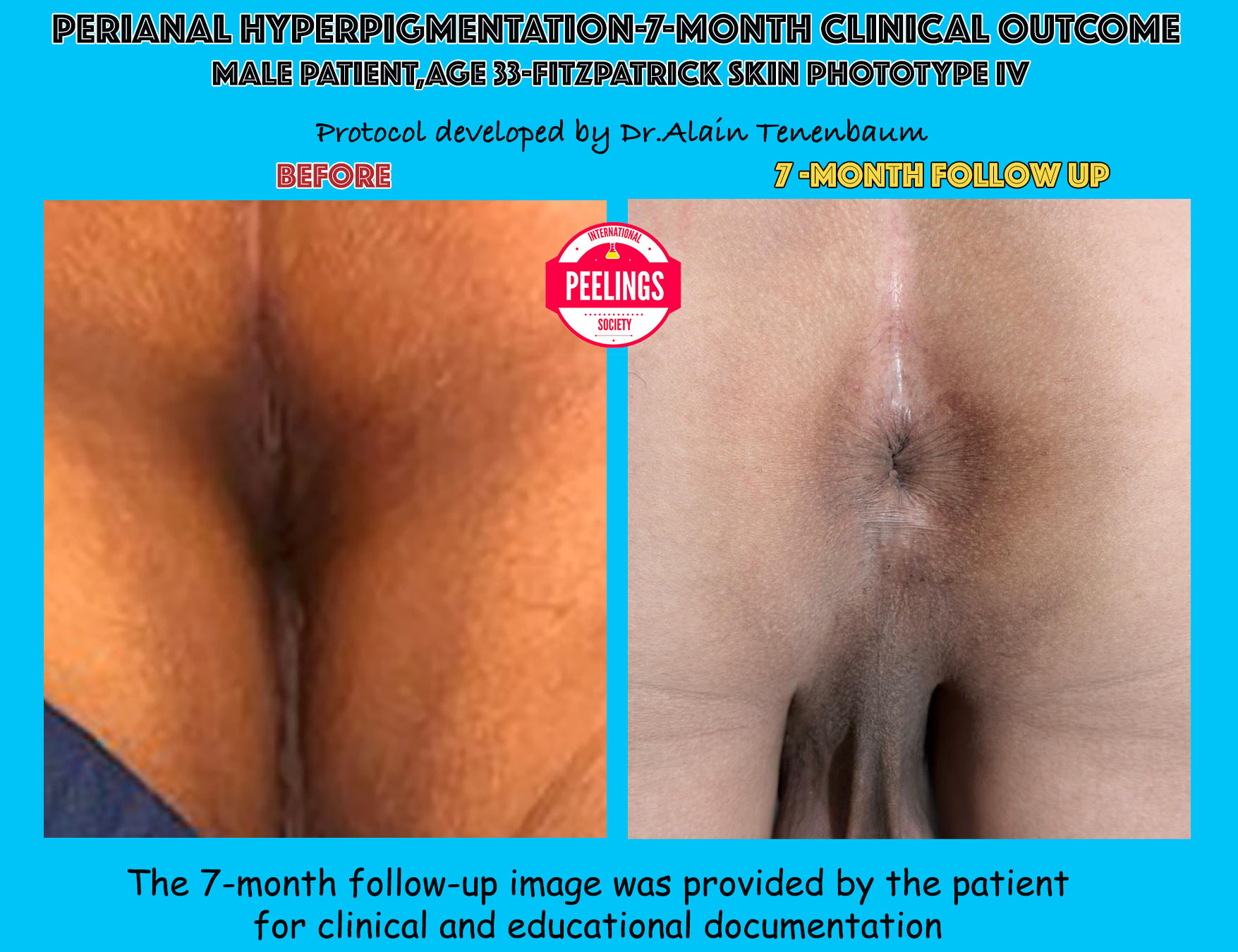
Case 01 · Documented Clinical Outcome

Perianal Hyperpigmentation

Baseline presentation compared with documented 7-month follow-up.

Patient Male
Age 33 Years
Phototype Fitzpatrick IV
Follow-Up 7 Months
Long-Term Clinical Documentation

Before → 7-Month Follow-Up

+ Zoom
Perianal hyperpigmentation clinical case showing baseline and 7-month follow-up in a 33-year-old male patient with Fitzpatrick skin phototype IV
Documented perianal hyperpigmentation outcome. Baseline appearance is compared with the 7-month follow-up photograph. The follow-up image was provided by the patient for clinical and educational documentation.
Clinical Reading

Long-Term Pigmentary Reassessment

The follow-up photograph documents the clinical appearance seven months after baseline. Its principal value is longitudinal: it allows the pigmentation pattern at baseline to be compared with the later documented state rather than judging response from an immediate post-procedure endpoint.

Endpoint ≠ Long-Term Outcome

Immediate whitening, pseudo-frosting, erythema or early desquamation describe procedural or early post-treatment events. They should not be confused with the final pigmentary result documented months later.

Case 01 · Recorded Treatment Course

Six Documented Treatment Sessions

The treatment calendar belongs to this documented clinical course and provides context for interpretation of the long-term follow-up. It is not presented as a universal schedule for every patient.

Clinical treatment calendar documenting six treatment sessions at approximately two to three week intervals
Documented case schedule. Six sessions with approximately 2–3 weeks between sessions, as recorded in the clinical documentation.

A Longitudinal Course — Not a Single Procedure

Baseline Initial perianal pigmentation documented before the treatment course.
Treatment Course Six recorded sessions separated by approximately 2–3 weeks in this individual case.
Long-Term Follow-Up Clinical appearance documented at seven months.
Case-specific schedule. Session number and interval should not be extrapolated automatically to another patient. Treatment planning remains dependent on anatomy, tissue response, pigmentary behaviour and clinical reassessment.
Clinical Case Timeline

Baseline → Treatment Course → Long-Term Outcome

Baseline Initial Pigmentation

Document the original distribution and clinical appearance.

›
Course Six Recorded Sessions

Reassessment occurs throughout a longitudinal treatment strategy.

›
Follow-Up 7-Month Documentation

Evaluate the evolved pigmentary state at long-term follow-up.

Interpretation of Clinical Images

Individual Outcome ≠ Guaranteed Outcome

Clinical photographs document the response of an individual patient under a specific treatment course. They do not establish a guaranteed degree of pigment reduction, a fixed number of sessions or an identical response in another patient. Anatomy, baseline pigmentation, phototype, inflammatory susceptibility and tissue response can all influence clinical evolution.

Next · Clinical Decision Matrix
From documented outcome to structured clinical decision-making.
Clinical Synthesis · 15

Clinical Decision Algorithm

The clinical pathway can be reduced to a structured sequence: define the anatomical field, confirm eligibility, identify the pigmentary problem, stratify inflammatory risk, select the therapeutic pathway and reassess the response.

From Anatomy to Therapeutic Pathway

This algorithm summarizes the decision logic developed throughout the preceding sections. It is a clinical synthesis rather than a replacement for assessment. Each decision remains dependent on the individual anatomical and pigmentary context.

Final Clinical Algorithm

Define → Exclude → Stratify → Select → Reassess

Each stage acts as a decision gate. Treatment proceeds only when the preceding condition has been clinically resolved.

01
Anatomical Gate

Define the Treatment Field

Is the pigmentation located on an anatomically identified external cutaneous surface appropriate for consideration as a treatment field?

Yes External Cutaneous Field Confirmed

Continue to clinical eligibility assessment.

No Transitional / Mucosal / Internal Tissue

Do not extend the external pigmentation protocol into the excluded anatomical field.

02
Eligibility Gate

Confirm That the Tissue Is Suitable for Treatment

Is the selected external cutaneous field clinically stable, intact and free from conditions requiring diagnosis, recovery or treatment before a pigmentation procedure?

Eligible Proceed to Pigmentary Analysis

The anatomical field is clinically suitable for further therapeutic decision-making.

Not Yet Eligible Stabilize / Diagnose / Reassess

Active inflammation, infection, barrier disruption, trauma or an undiagnosed lesion takes priority.

03
Pigmentary Gate

Identify the Dominant Pigmentary Problem

Interpret the visible pigmentation within the metabolic pigment model rather than treating colour alone.

Compartment 01 Melanin Production
Compartment 02 Melanin Transport
Compartment 03 Pigment Processing & Clearance
04
Risk Modifier

Stratify PIH Susceptibility

Integrate phototype with previous PIH, inflammatory history, friction, current tissue condition and the inflammatory potential of the proposed intervention.

Lower Concern Standard Clinical Caution
Intermediate Concern Increase Procedural Restraint
Higher Concern Prioritize Pigmentary Control
05
Therapeutic Gate

Select the Therapeutic Pathway

Match the treatment objective to the anatomical field, pigmentary state, tissue tolerance and PIH susceptibility.

Pathway A
Controlled Correction

Consider a corrective approach only when clinically indicated and compatible with the selected external cutaneous field and individual inflammatory risk.

or
Pathway B
Metabolic Regulation

Prioritize progressive pigmentary regulation when the clinical objective, tissue condition or risk profile favours a less corrective strategy.

Step 06 · Mandatory Reassessment

Treat the Evolved Clinical State

After each treatment phase, reassess tissue recovery, pigmentation and inflammatory response. The next intervention should be based on the current clinical state rather than on the previous session or a predetermined sequence.

Longitudinal Strategy

The Pathway Changes as Pigmentation Changes

Phase 01 Correction

Reduce clinically relevant visible pigmentation when corrective treatment is indicated.

›
Phase 02 Stabilization

Consolidate the response while progressively reducing unnecessary inflammatory stimulus.

›
Phase 03 Maintenance

Preserve pigmentary stability through an increasingly regulatory strategy.

Clinical Decision Rule

Anatomy First. Eligibility Second. Mechanism Before Intervention.

A therapeutic pathway should never be selected from pigmentation colour alone. Anatomical tissue type determines where treatment may occur; clinical eligibility determines whether it should occur; pigmentary behaviour and PIH susceptibility determine how cautiously the strategy should proceed.

Next · Related Protocols & Products
Continue from clinical decision-making to the relevant therapeutic resources.
Clinical Resources · 16

Continue the Clinical Pathway

Continue from the clinical framework for intimate hyperpigmentation to related treatment protocols, product resources and complementary clinical topics. These resources provide deeper guidance without repeating the decision process developed on this page.

→
Clinical Navigation

Select the next resource according to what you need: a practical protocol, a product-specific resource, or additional clinical context.

Need Procedure Detail? Clinical Protocols
›
Need Product Detail? Product Resources
›
Need Broader Context? Clinical Topics
Resource Navigation Principle

Move Deeper Without Repeating the Clinical Decision Process

The resources above extend specific components of the intimate hyperpigmentation framework. They should be read according to the clinical question being addressed: regional procedure, product knowledge or broader pigmentary context.

Next · Frequently Asked Questions
Practical answers to recurring clinical questions.
Clinical FAQ · 17

Frequently Asked Clinical Questions

Practical answers to recurring questions about anatomy, pigmentation, treatment selection, clinical endpoints, PIH risk and long-term management in intimate hyperpigmentation.

Anatomy Treatment Safety Pigmentation Follow-Up
01 Anatomy Can all pigmented intimate areas be treated in the same way?

No. Anatomical location alone does not define tissue type. External cutaneous surfaces, transitional epithelial structures and internal mucosal tissues are not therapeutically interchangeable.

Treatment must therefore begin by defining the actual anatomical treatment field before selecting any corrective or regulatory approach.

External anatomical location does not automatically mean external cutaneous tissue.
02 Anatomical Safety Should the labia minora or vulvar vestibule be included in the treatment field?

No. Within the clinical framework described on this page, the labia minora, vulvar vestibule and internal mucosal structures are anatomical do-not-treat zones.

A procedure intended for external cutaneous tissue should not simply be extended medially because visible pigmentation continues beyond the cutaneous field.

Pigment distribution does not override anatomical boundaries.
03 Anatomical Landmark Does the presence or absence of hair define the vulvar treatment boundary?

Hair-bearing skin can be a useful practical anatomical landmark, particularly when identifying external labia majora skin, but it is not a histological definition by itself.

The clinically relevant distinction remains the transition between external cutaneous tissue and transitional or internal epithelial structures.

Hair distribution is a clue — not the definition of tissue type.
04 Pigmentation Is intimate hyperpigmentation a single uniform condition?

No. Similar visible pigmentation may arise in different clinical contexts, including repetitive friction, post-inflammatory pigmentation, hormonal influence, constitutional pigmentary tendency and repeated external irritation.

The objective is therefore not simply to identify how dark the area appears, but to understand the dominant mechanisms maintaining the pigmentary state.

Treat the pigmentary mechanism — not colour alone.
05 Therapeutic Strategy Does every patient require a corrective acid-based procedure?

No. The therapeutic pathway depends on the anatomical field, tissue condition, pigmentary state, clinical objective and individual susceptibility to post-inflammatory hyperpigmentation.

Some selected external cutaneous fields may justify a controlled corrective pathway, whereas other situations favour a predominantly metabolic regulatory approach.

Corrective intensity should be clinically selected — never automatic.
06 Treatment Sequence Where does Peeling de Luxe Plus belong when an acid-based corrective step is used?

Within the treatment framework described here, Peeling de Luxe Plus is positioned after the selected corrective acid step and before subsequent metabolic products.

Corrective Acid → Clinical Endpoint → Peeling de Luxe Plus → Metabolic Step
07 Clinical Endpoint Is visible frosting the objective of treatment?

No. A visible endpoint may provide clinical information during a selected corrective procedure, but it should not become an objective that is pursued independently of tissue response.

The clinician should observe the endpoint rather than chase it. A more visible immediate reaction does not by itself establish a better long-term result.

Clinical Endpoint ≠ Clinical Outcome
08 PIH Risk Can PIH risk be predicted from Fitzpatrick phototype alone?

No. Phototype contributes useful information, but PIH susceptibility should be interpreted as a combined clinical profile.

Previous PIH, active or recurrent inflammation, friction, recent trauma, current barrier condition and the inflammatory potential of the proposed intervention all contribute to the risk assessment.

Stratify the patient — not the phototype alone.
09 Treatment Course Is there a fixed number of sessions for intimate hyperpigmentation?

No universal session number should be inferred from an individual clinical case. Treatment duration depends on baseline pigmentation, anatomical site, mechanism, response, recovery and pigmentary stability.

The clinical state should be reassessed over time rather than forcing every patient into a predetermined treatment calendar.

Treat the evolved clinical state — not the previous session.
10 Long-Term Strategy Does treatment end when the pigmentation becomes visibly lighter?

Not necessarily. Visible correction represents only one stage of the longitudinal strategy. Once sufficient correction has been achieved, the therapeutic emphasis can shift toward stabilization and maintenance.

The objective is not repeated correction indefinitely, but progressive control of the pigmentary state with less unnecessary inflammatory stimulus.

Correction → Stabilization → Maintenance
11 Contraindications When should treatment be postponed rather than performed?

Treatment should be deferred when the proposed field presents active inflammation, suspected infection, erosion or ulceration, significant barrier disruption, recent local trauma or procedure, or an undiagnosed lesion requiring evaluation.

In such circumstances, diagnosis, recovery or stabilization takes priority over pigment correction.

Eligibility comes before intervention.
12 Clinical Outcome Can the result of one documented before-and-after case be expected in every patient?

No. Clinical photographs document the response of an individual patient under a particular clinical context. They do not establish a guaranteed degree of pigment reduction, a fixed treatment duration or an identical response in another patient.

Outcomes should always be interpreted in relation to anatomical area, baseline pigmentation, treatment course, tissue response and follow-up interval.

Individual Outcome ≠ Guaranteed Outcome
Clinical Take-Home

The Same Visible Pigmentation Does Not Always Require the Same Treatment

Intimate hyperpigmentation should be approached through anatomical identification, clinical eligibility, mechanism-based interpretation, PIH risk stratification and longitudinal reassessment. The safest therapeutic decision is determined by the tissue and its clinical state, not by colour alone.

Next · References & Professional Information
Scientific context, clinical documentation and professional use.
Scientific Authorship · 18

Scientific Publications & Author Contributions

Selected published contributions by Alain Tenenbaum, M.D., Ph.D., D.Sc., documenting the scientific development of concepts related to chemical peel chemistry, mechanisms of action and clinical classification.

AT
Selected Scientific Contributions Alain Tenenbaum, M.D., Ph.D., D.Sc.
Peel Chemistry Mechanisms Classification Clinical Concepts
01 Book Chapter
Chemical Peels, Procedures in Cosmetic Dermatology Series, second edition
Chemical Peels · 2nd Edition

Chemistry of Peelings and Hypothesis of the Mechanism of Action

Chapter Authors Luc Dewandre · Alain Tenenbaum

Published contribution addressing the chemistry of chemical peeling and a mechanistic interpretation of peel action.

Procedures in Cosmetic Dermatology Series Saunders / Elsevier 2011
Chemistry Mechanism of Action
02 Book Chapter
Chemical Peels, Procedures in Cosmetic Dermatology Series, third edition
Chemical Peels · 3rd Edition

The Chemistry of Peels: A Hypothesis of Mechanism of Action and Classification of Peels

Chapter Authors Luc Dewandre · Alain Tenenbaum · Desmer Destang

A continued scientific contribution integrating peel chemistry, mechanisms of action and chemical peel classification.

Procedures in Cosmetic Dermatology Series Edited by Suzan Obagi
Mechanism Classification Metabolic Interactions
03 International Contribution
Face In Harmony, A Rejuvenation Encyclopedia Across the Globe
International Collective Work

Face In Harmony A Rejuvenation Encyclopedia Across the Globe

Author Contribution Alain Tenenbaum · Contributing Author

Contribution to the international reference work coordinated by Prof. Múcio Porto and contributing authors in the field of facial rejuvenation and aesthetic medicine.

Prof. Múcio Porto & Co-Authors International Expert Contribution
Facial Rejuvenation Aesthetic Medicine
Scientific Continuity
From Published Concepts to Contemporary Clinical Models

Published work on peel chemistry, mechanisms of action and classification forms part of the scientific background from which later clinical concepts have evolved.

The contemporary models presented on ChemicalPeeling.com extend this work into mechanism-driven clinical frameworks integrating tissue response, pigment regulation and metabolic interactions.

≠

Published contribution and contemporary clinical framework are distinct levels of scientific information. Current clinical models should not be interpreted as identical to, or automatically validated by, earlier published material.

Medical Documentation Explore Scientific Publications
View Publications
Clinical Continuation · 19
From Clinical Understanding

Continue Into Treatment Strategy.

Intimate hyperpigmentation requires anatomical precision, mechanism-driven selection and controlled pigmentary management. Continue with the clinical protocols and treatment resources used to translate these principles into practice.

01 Define Anatomy
02 Identify Mechanism
03 Select Strategy
04 Reassess Response
+
Need Product Guidance? Clinical Product Selection Matrix

Compare products according to clinical indication, therapeutic role and mechanism-driven treatment logic.

Open Selection Matrix
Clinical Principle

Anatomy first. Mechanism before intervention. Response before repetition.

Intimate Hyperpigmentation
Mechanism-Driven Clinical Strategy

Share this page with your network